. 2023 Oct;36(5):2125-2137.
doi: 10.1007/s10278-023-00866-1. Epub 2023 Jul 5.

Artificial Intelligence Application to Screen Abdominal Aortic Aneurysm Using Computed tomography Angiography

Affiliations

Artificial Intelligence Application to Screen Abdominal Aortic Aneurysm Using Computed tomography Angiography

Giovanni Spinella et al. J Digit Imaging. 2023 Oct.

Abstract

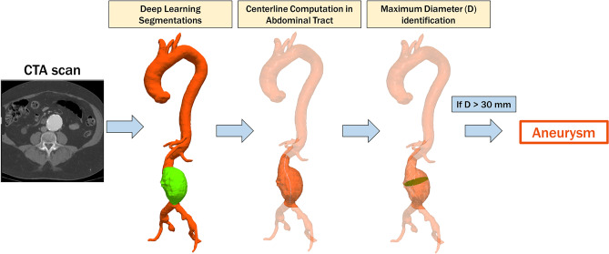
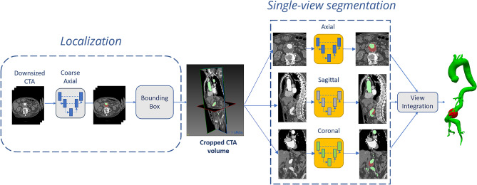
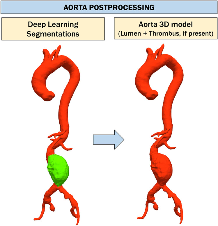
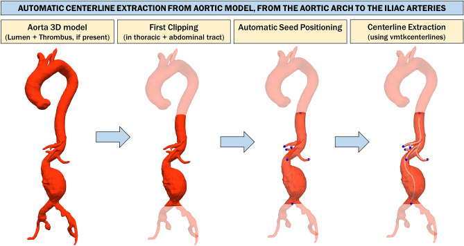
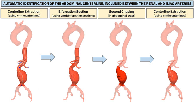
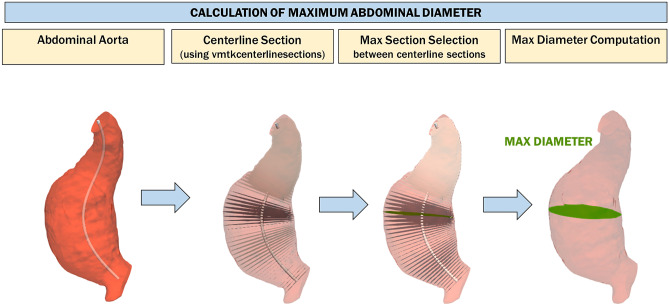
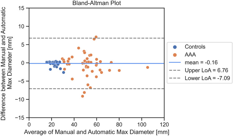
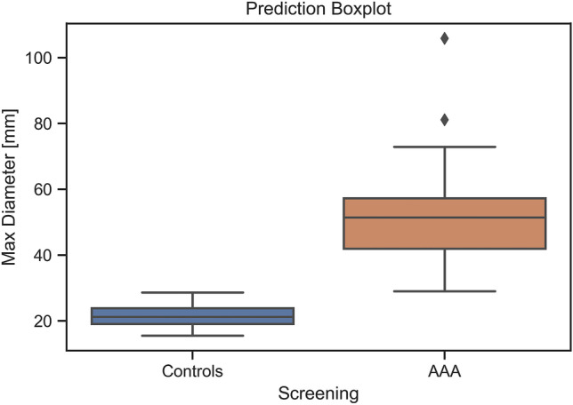
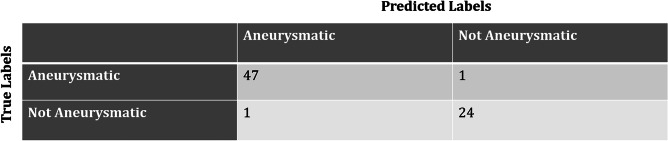
The aim of our study is to validate a totally automated deep learning (DL)-based segmentation pipeline to screen abdominal aortic aneurysms (AAA) in computed tomography angiography (CTA) scans. We retrospectively evaluated 73 thoraco-abdominal CTAs (48 AAA and 25 control CTA) by means of a DL-based segmentation pipeline built on a 2.5D convolutional neural network (CNN) architecture to segment lumen and thrombus of the aorta. The maximum aortic diameter of the abdominal tract was compared using a threshold value (30 mm). Blinded manual measurements from a radiologist were done in order to create a true comparison. The screening pipeline was tested on 48 patients with aneurysm and 25 without aneurysm. The average diameter manually measured was 51.1 ± 14.4 mm for patients with aneurysms and 21.7 ± 3.6 mm for patients without aneurysms. The pipeline correctly classified 47 AAA out of 48 and 24 control patients out of 25 with 97% accuracy, 98% sensitivity, and 96% specificity. The automated pipeline of aneurysm measurements in the abdominal tract reported a median error with regard to the maximum abdominal diameter measurement of 1.3 mm. Our approach allowed for the maximum diameter of 51.2 ± 14.3 mm in patients with aneurysm and 22.0 ± 4.0 mm in patients without an aneurysm. The DL-based screening for AAA is a feasible and accurate method, calling for further validation using a larger pool of diagnostic images towards its clinical use.

Keywords: Abdominal aortic aneurysm (AAA); Artificial intelligence (AI); Deep learning (DL); Screening.


Conflict of interest statement

The authors have no relevant financial or non-financial interests to disclose.

Figures

Fig. 1
Fig. 1
Pipeline proposed to perform automatic aneurysm screening. First, aortic lumen and thrombus are extracted from the CTA scan with a deep learning algorithm. Then, the lumen centerline is computed in the abdominal tract. Finally, the maximum abdominal aortic diameter is extracted. If the diameter is greater than the threshold value, the patient is considered “aneurysmatic”
Fig. 2
Fig. 2
Pipeline performing automatic lumen segmentation from CTA images. A first network is used to localize the region of interest (ROI) containing the aorta (in green); then, the ROI is processed by three orthogonal networks. The predictions are integrated to obtain a final segmentation. The same pipeline is adopted to segment the intraluminal thrombus (in red) from CTA scans
Fig. 3
Fig. 3
Aorta post-processing step. First, aortic segmentation is obtained by a summation operation between the segmentation of the aortic lumen and the thrombus, respectively. Then, this segmentation undergoes a smoothing process exploited to eliminate irregularities that can adversely affect the next steps. Finally, the polygonal model of aorta (lumen + thrombus, if present) is obtained using the marching cube algorithm [31]
Fig. 4
Fig. 4
Automatic centerline extraction from aortic model, from the aortic arch to the iliac arteries. The 3D model is cut between z-coordinates related to the aortic arch and iliac arteries, which are identified through a process of connected component analysis performed slice by slice on the axial view. A raw centerline is obtained from this model using a skeletonization algorithm [29]. The skeleton is exploited to automatically extract centerline end-point voxels that will be used to calculate a more refined centerline with the vmtkcenterlines function of Vascular Modeling ToolKit (VMTK) [30] on the aorta 3D model
Fig. 5
Fig. 5
Automatic identification of the abdominal centerline, included between renal and iliac arteries. The surface and relative centerlines, both of which have already been divided into branches (using the vmtkbranchextractor and vmtkbranchclipper functions), are used to calculate the bifurcation sections of the surface using the vmtkbifurcationsections function. The last bifurcation plane extracted from the 3D model is always relative to the renal arteries. The aortic model is clipped in the abdominal area using two planes perpendicular to the centerline, centered at the height of the superior renal artery and at the height of the iliac bifurcation, respectively. The origins of these planes are used as the source seed and target seed, respectively, to recalculate the centerline of the isolated abdominal tract
Fig. 6
Fig. 6
Calculation of the maximum abdominal diameter. Sections perpendicular to the centerline are generated with the function vmtkcenterlinesections. The diameter of each section is computed, and the section with the largest diameter is selected among them all
Fig. 7
Fig. 7
Bland–Altman plot
Fig. 8
Fig. 8
Box plot representing maximum diameters predicted in patients affected by AAA and controls
Fig. 9
Fig. 9
Confusion matrix

References

    1. Wanhainen A, Verzini F, Van Herzeele I, Allaire E, Bown M, Cohnert T, Dick F, van Herwaarden J, Karkos C, Koelemay M, Kölbel T, Loftus I, Mani K, Melissano G, Powell J, Szeberin Z. Esvs Guidelines Committee, de Borst GJ, Chakfe N, Debus S, Verhagen H: Editor's Choice - European Society for Vascular Surgery (ESVS) 2019 Clinical Practice Guidelines on the Management of Abdominal Aorto-iliac Artery Aneurysms. European journal of vascular and endovascular surgery: the official journal of the European Society for Vascular Surgery. 2019;57(1):8–93. doi: 10.1016/j.ejvs.2018.09.020. - DOI - PubMed
    1. Chaikof EL, Dalman RL, Eskandari MK, Jackson BM, Lee WA, Mansour MA, Mastracci TM, Mell M, Murad MH, Nguyen LL, Oderich GS, Patel MS, Schermerhorn ML, Starnes BW: The Society for Vascular Surgery practice guidelines on the care of patients with an abdominal aortic aneurysm. Journal of vascular surgery, 67(1), 2–77.e2, 2018.Cosford, PA, Leng GC: Screening for abdominal aortic aneurysm. The Cochrane Database of Systematic Reviews, (2), CD002945, 2007 - PubMed
    1. Sonesson B, Lanne T, Hansen F, Sandgren T. Infrarenal aortic diameter in the healthy person. Eur J Vasc Surg. 1994;8(1):89–95. doi: 10.1016/S0950-821X(05)80127-6. - DOI - PubMed
    1. Altobelli E, Rapacchietta L, Profeta VF, Fagnano R. Risk Factors for Abdominal Aortic Aneurysm in Population-Based Studies: A Systematic Review and Meta-Analysis. Int J Environ Res Public Health. 2018;15(12):2805. doi: 10.3390/ijerph15122805. - DOI - PMC - PubMed
    1. O’Leary SA, Kavanagh EG, Grace PA, McGloughlin TM, Doyle BJ. The biaxial mechanical behavior of abdominal aortic aneurysm intraluminal thrombus: classification of morphology and the determination of layer and region specific properties. J Biomech. 2014;47(6):1430–1437. doi: 10.1016/j.jbiomech.2014.01.041. - DOI - PubMed

Publication types

MeSH terms

LinkOut - more resources