. 2023 Jul 5;21(1):437.
doi: 10.1186/s12967-023-04208-1.

Intraparenchymal convection enhanced delivery of AAV in sheep to treat Mucopolysaccharidosis IIIC

Affiliations

Intraparenchymal convection enhanced delivery of AAV in sheep to treat Mucopolysaccharidosis IIIC

Claire O'Leary et al. J Transl Med. .

Abstract

Background: Mucopolysaccharidosis IIIC (MPSIIIC) is one of four Sanfilippo diseases sharing clinical symptoms of severe cognitive decline and shortened lifespan. The missing enzyme, heparan sulfate acetyl-CoA: α-glucosaminide-N-acetyltransferase (HGSNAT), is bound to the lysosomal membrane, therefore cannot cross the blood-brain barrier or diffuse between cells. We previously demonstrated disease correction in MPSIIIC mice using an Adeno-Associated Vector (AAV) delivering HGSNAT via intraparenchymal brain injections using an AAV2 derived AAV-truetype (AAV-TT) serotype with improved distribution over AAV9.

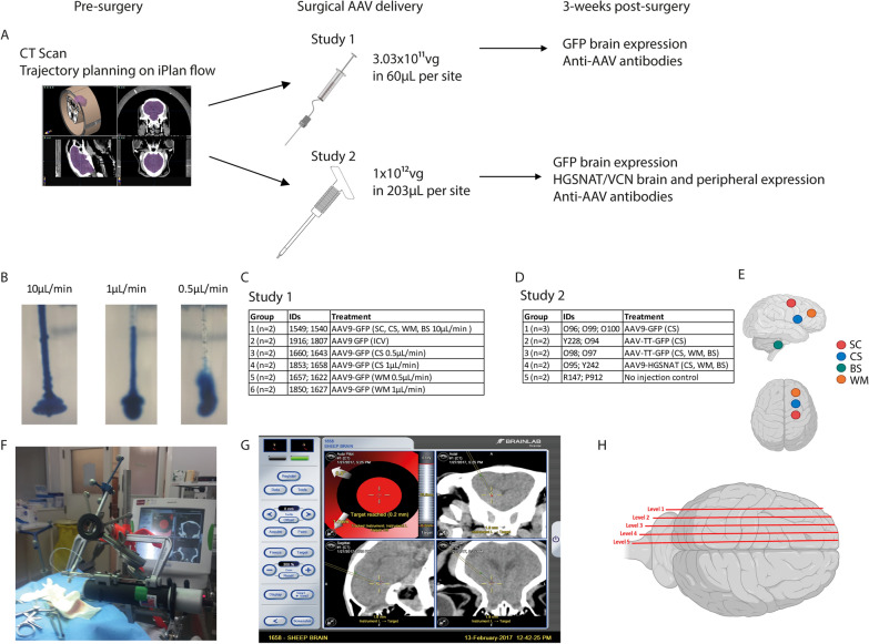
Methods: Here, intraparenchymal AAV was delivered in sheep using catheters or Hamilton syringes, placed using Brainlab cranial navigation for convection enhanced delivery, to reduce proximal vector expression and improve spread.

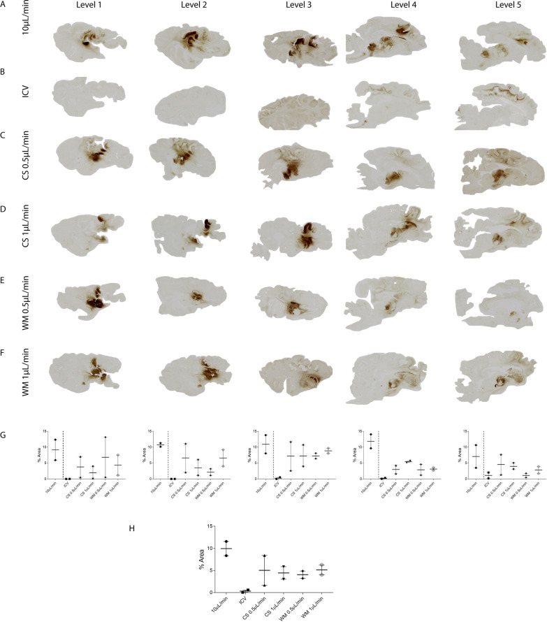
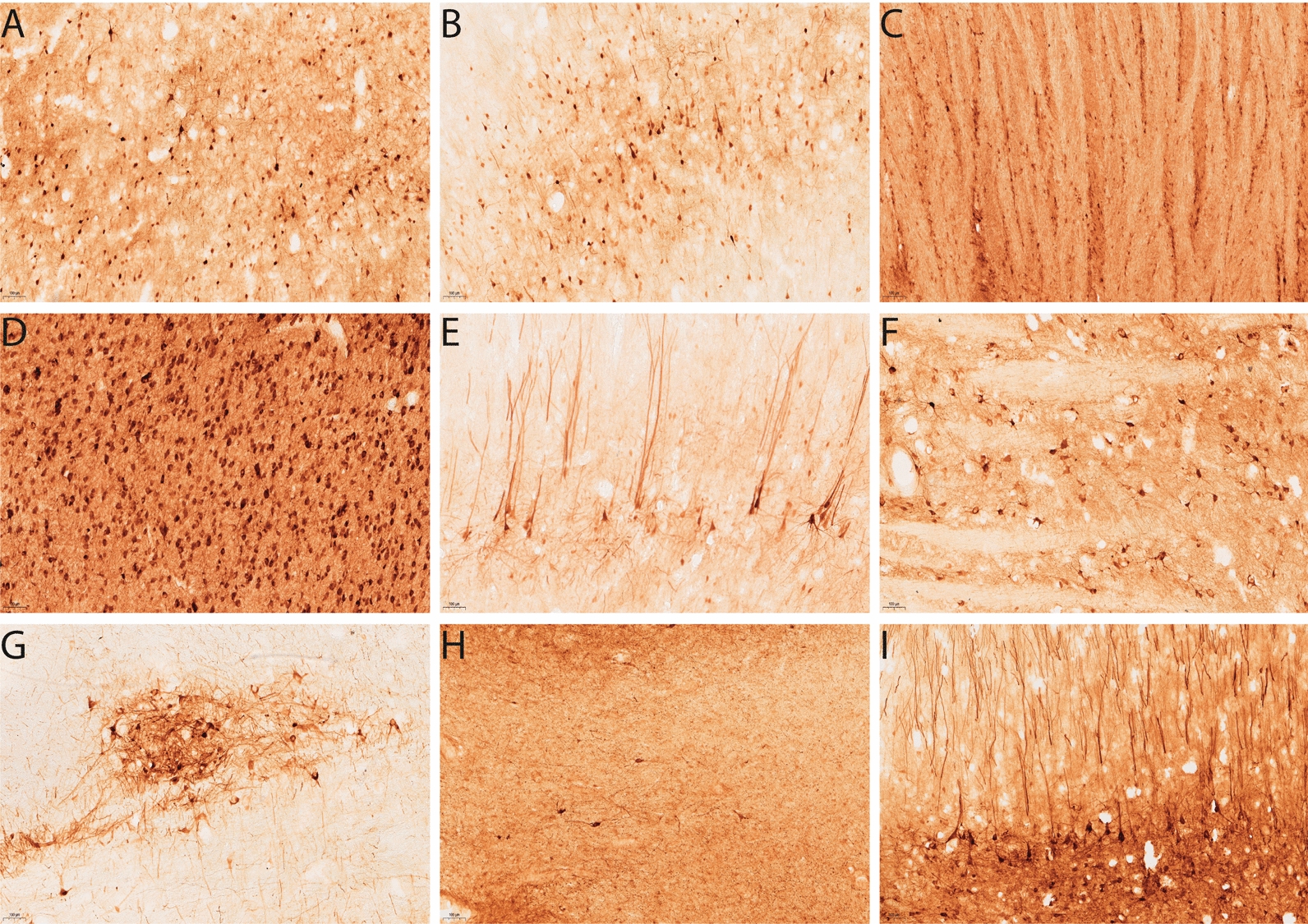
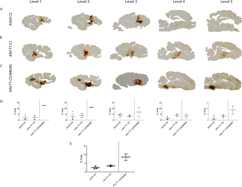
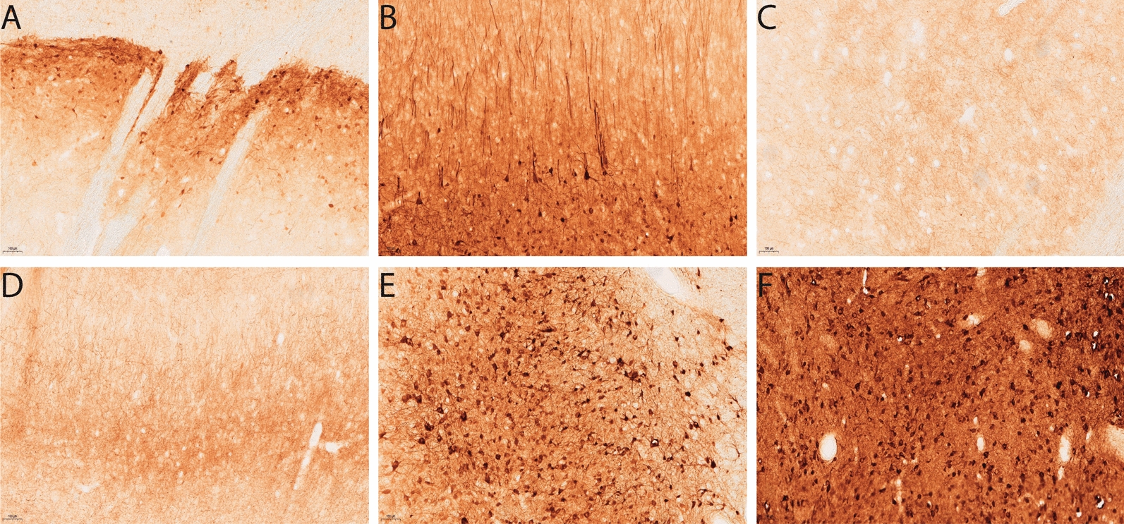
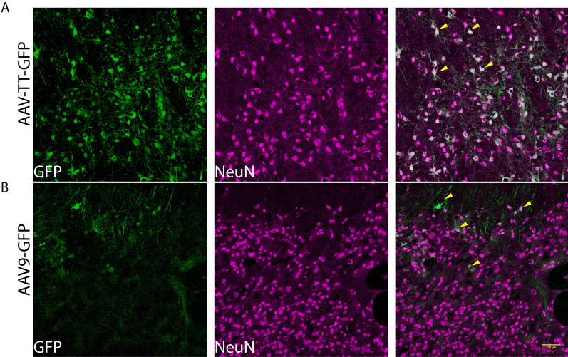
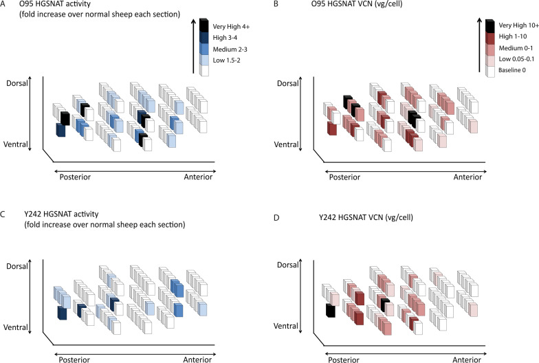
Results: Hamilton syringes gave improved AAV-GFP distribution, despite lower vector doses and titres. AAV-TT-GFP displayed moderately better transduction compared to AAV9-GFP but both serotypes almost exclusively transduced neurons. Functional HGSNAT enzyme was detected in 24-37% of a 140g gyrencephalic sheep brain using AAV9-HGSNAT with three injections in one hemisphere.

Conclusions: Despite variabilities in volume and titre, catheter design may be critical for efficient brain delivery. These data help inform a clinical trial for MPSIIIC.

Keywords: AAV gene therapy; Convection Enhanced Delivery; HGSNAT; Large animal; Mucopolysaccharidosis.


Conflict of interest statement

Delivery of HGSNAT using AAV to MPSIIIC is the subject of a patent held by BB, COL and EH. BB is a consultant and grant holder for AVROBIO, Orchard Therapeutics and Phoenix Nest. BB and COL received payments from Phoenix Nest for previous MPSIIIC work.

Figures

Fig. 1.
Fig. 1.
Experimental protocol for AAV distribution studies. A Overview of treatment protocol and tissue analysis. B Different flow rates in 0.4% agarose gel phantoms. Experimental groups used in study 1 C and study 2 D. E Schematic of injection sites for Corpus Striatum (CS), White Matter (WM), Subcortical zone (SC) and Brainstem (BS) from a lateral and superior view. F Syringe driver used for AAV delivery. G Example surgical screenshot from Brainlab showing target accuracy. H Schematic of mediolateral sagittal brain levels 1-5 used for immunohistochemical analysis
Fig. 2
Fig. 2
Comparative distribution of GFP expression in sheep brains in study 1. Images showing GFP expression in sheep brains 3 weeks post-injection. Sheep received AAV9-GFP (n=2/group) either into A four locations: the corpus striatum (CS), white matter (WM), subcortical zone (SC) and brainstem (BS), B the lateral ventricles (ICV), (C, D) CS and (E, F) WM with the latter two groups separated into two infusion flow rates (0.5 µL/min and 1 μL/min). G Average GFP expression across each sagittal brain level. H Average GFP expression across the entire hemisphere. Data are presented as mean ± s.e.m
Fig. 3
Fig. 3
GFP expression from different injection sites in sheep brains in study 1. Images revealing the pattern of GFP expression 3 weeks post AAV9-GFP injection at 10× magnification. After delivery into the lateral ventricles transduced cells can be visualised on the A dorsal and B ventral surface of the ventricle. GFP transduced cells can be visualised in the C striatum, D medial geniculate nucleus , E motor cortex, F brainstem and G thalamus after direct injection into the CS. For WM injections, GFP positivity was detected in the H WM and the I neurons adjacent to the injection site. Scale bar = 100 µm
Fig. 4
Fig. 4
Comparative distribution of GFP expression in sheep brains in study 2. Images showing GFP expression in sheep brains 3 weeks post-injection. Two groups received either A AAV9-GFP (n=3) or B AAV-TT GFP (n=2) into the corpus striatum (CS) only. C An additional group of sheep (n=2) received AAV-TT-GFP into the CS, white matter (WM) and brainstem (BS). D Average GFP expression across each sagittal brain level. E Average GFP expression across the entire hemisphere. Data are presented as mean ± s.e.m
Fig. 5
Fig. 5
GFP expression from serotypes in sheep brains in study 2. Images revealing the pattern of GFP expression 3 weeks post AAV-GFP injection at 10× magnification. AAV9-GFP injection into the corpus striatum (CS) displays GFP transduced cells in the A striatum and B motor cortex. AAV-TT-GFP transduced cells can be visualised in the C striatum, D motor cortex, E medial geniculate nucleus and F thalamus. Scale bar = 100 µm
Fig. 6
Fig. 6
AAV vectors target neurons in the corpus striatum of the sheep brain. Double staining of GFP+ (green) with NeuN+ neurons (magenta) in A AAV-TT-GFP and B AAV9-GFP treated sheep at 10× magnification. Arrows in the merged image indicate examples of co-localization of GFP+ and NeuN+ cells. Scale bar = 100 µm
Fig. 7
Fig. 7
Immune reactions pre- and post-injection. Total IgG antibody responses against AAV9 and AAV-TT capsid proteins measured by ELISA on day 0 (the day of surgery) and 3 weeks post-surgery (at necropsy) in study 1 (A, B) and study 2 (CJ). A number of sheep showed pre-existing antibodies to the AAV-capsids that were still evident at necropsy. D One sheep developed an immune response to the AAV capsid. J One sheep (highlighted in red) was sacrificed on the day of surgery due to an adverse event but had no IgG antibodies against the capsid
Fig. 8
Fig. 8
Comparison of HGSNAT expression in AAV9-HGSNAT-treated sheep 3 weeks post-surgery. Vector distribution is represented by three-dimensional schematic drawings of the brain. Each brain was divided into 1cm3 cubes. Quantitative comparison of HGSNAT activity above endogenous background level was assessed (A, C). HGSNAT values were determined as fold increase compared to the mean of 2 un-injected sheep for that relative brain section and VCN in two different sheep O95 and Y242 (C, D)

References

    1. Fedele AO. Sanfilippo syndrome: causes, consequences, and treatments. Appl Clin Genet. 2015;8:269–81. doi: 10.2147/TACG.S57672. - DOI - PMC - PubMed
    1. Ghosh A, et al. Recommendations on clinical trial design for treatment of Mucopolysaccharidosis Type III. Orphanet J Rare Dis. 2017;12(1):117. doi: 10.1186/s13023-017-0675-4. - DOI - PMC - PubMed
    1. Tordo J, et al. A novel adeno-associated virus capsid with enhanced neurotropism corrects a lysosomal transmembrane enzyme deficiency. Brain. 2018;141(7):2014–2031. doi: 10.1093/brain/awy126. - DOI - PMC - PubMed
    1. Tardieu M, et al. Intracerebral administration of adeno-associated viral vector serotype rh.10 carrying human SGSH and SUMF1 cDNAs in children with mucopolysaccharidosis type IIIA disease: results of a phase I/II trial. Human Gene Ther. 2014;25(6):506–16. doi: 10.1089/hum.2013.238. - DOI - PubMed
    1. Tardieu M, et al. Intracerebral gene therapy in children with mucopolysaccharidosis type IIIB syndrome: an uncontrolled phase 1/2 clinical trial. Lancet Neurol. 2017;16(9):712–720. doi: 10.1016/S1474-4422(17)30169-2. - DOI - PubMed

Publication types

LinkOut - more resources