. 2023 Jul 5;22(1):104.
doi: 10.1186/s12943-023-01806-x.

A novel polypeptide CAPG-171aa encoded by circCAPG plays a critical role in triple-negative breast cancer

Affiliations

A novel polypeptide CAPG-171aa encoded by circCAPG plays a critical role in triple-negative breast cancer

Runjie Song et al. Mol Cancer. .

Abstract

Background: The treatment of Triple-negative breast cancer (TNBC) has always been challenging due to its heterogeneity and the absence of well-defined molecular targets. The present study aims to elucidate the role of protein-coding circRNAs in the etiology and carcinogenesis of TNBC.

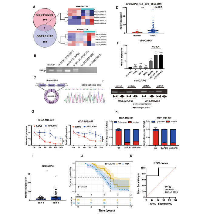
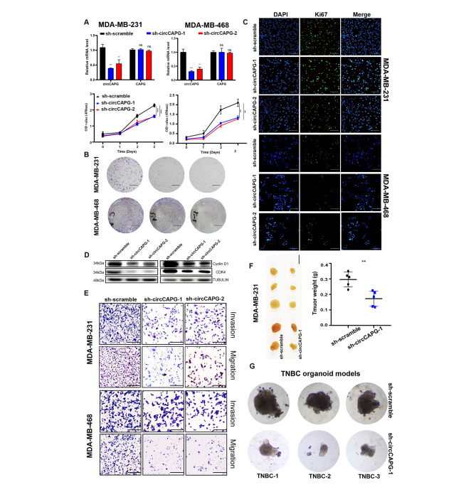
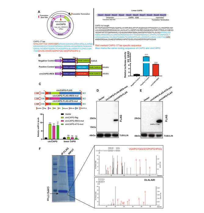
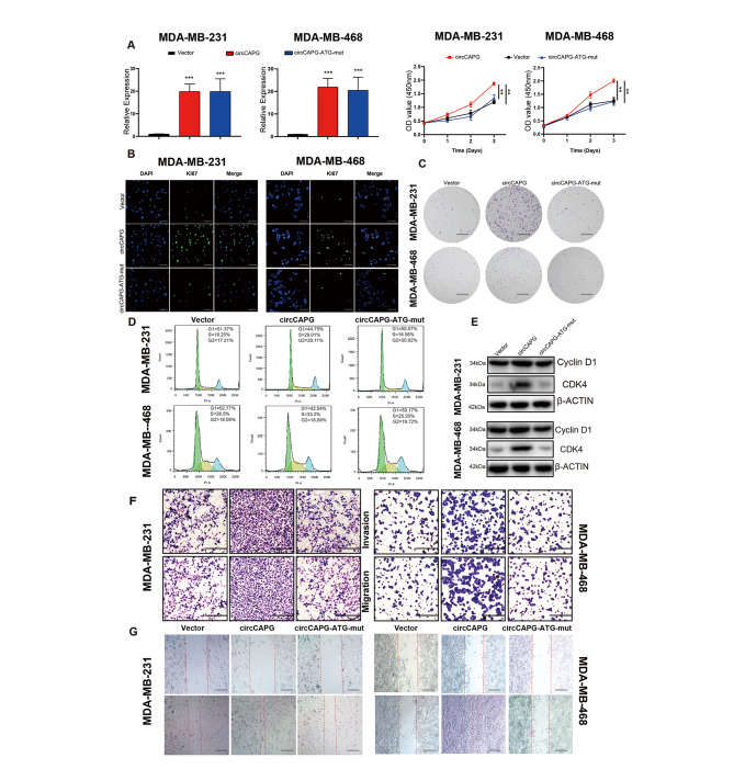
Methods: CircRNA expression data in TNBC (GEO: GSE113230, GSE101123) were reanalyzed and then circCAPG was selected for further study. To identify the polypeptide-coding function of circCAPG, a series of experiments, such as Mass spectrometry and dual-luciferase reporter assays were conducted. Cell proliferation, apoptosis and metastasis parameters were determined to investigate the cancerous functions CAPG-171aa plays in both TNBC organoids and nude mice. Mechanistically, the relation between CAPG-171aa and STK38 in TNBC was verified by immunoprecipitation analyses and mass spectrometry. The interactions between SLU7 and its binding site on circCAPG were validated by RIP-qPCR experiments.

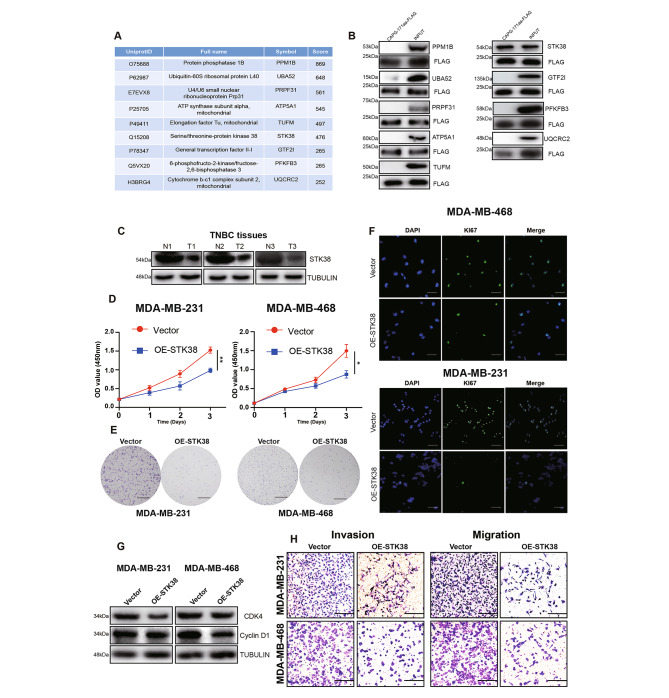
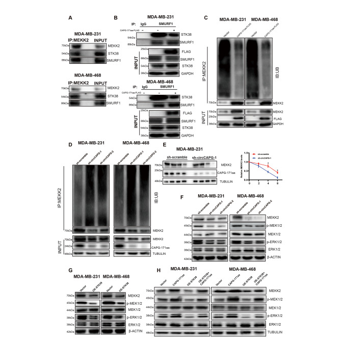
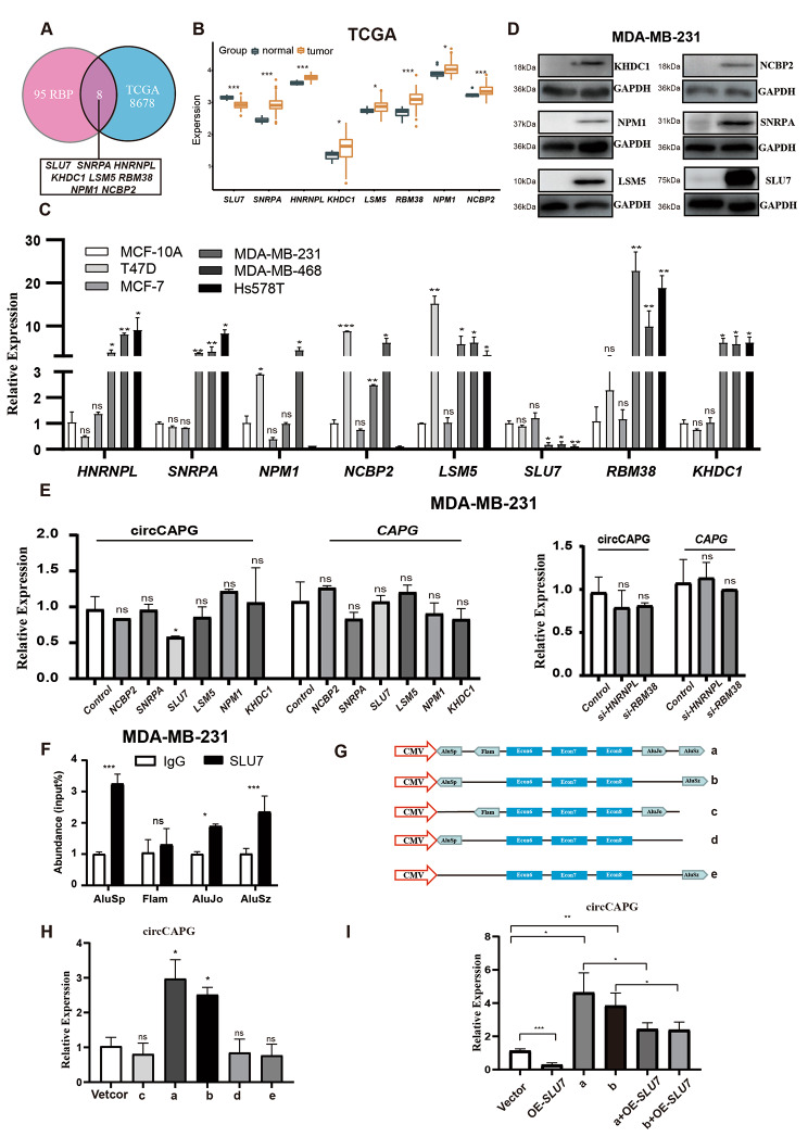
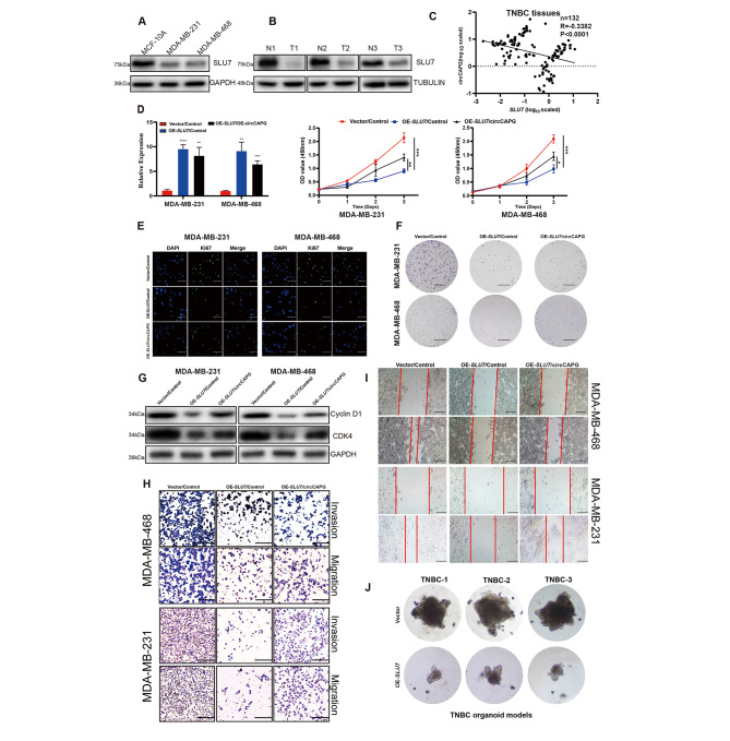
Results: In both TNBC clinical samples and cell lines, the expression level of circCAPG was identified to be higher compared with normal ones and positively correlated with the overall survival (n = 132) in a 10-year follow-up study, in which the area under the curve of receiver operating characteristic was 0.8723 with 100% specificity and 80% sensitivity. In addition, we found that circCAPG knockdown (KD) significantly inhibited the growth of TNBC organoids. Intriguingly, circCAPG can be translated into a polypeptide named CAPG-171aa which promotes tumor growh by disrupting the binding of serine/threonine kinase 38 (STK38) to SMAD-specific E3 ubiquitin protein ligase 1 (SMURF1) and thereby preventing MEKK2 ubiquitination and proteasomal degradation. Furthermore, we found that SLU7 Homolog- Splicing Factor (SLU7) can regulate the bio-generation of circCAPG through binding to the flanking Alu sequences of circRNA transcripts.

Conclusions: circCAPG significantly enhances the proliferation and metastasis of TNBC cells by encoding a novel polypeptide CAPG-171aa and afterwards activates MEKK2-MEK1/2-ERK1/2 pathway. Additionally, the formation of circCAPG is found to be mediated by SLU7. The present study provides innovative insight into the role of protein-coding circRNAs CAPG-171aa in TNBC, and its capacity to serve as a promising prognostic biomarker and potential therapeutic target in TNBC.

Keywords: CAPG-171aa; CircCAPG; MEKK2; SLU7; STK38; TNBC; Therapeutic target.


Conflict of interest statement

All authors declare that they have no competing interests.

Figures

Fig. 1
Fig. 1
CircCAPG is highly expressed in TNBC. (A) Differentially expressed circRNAs in TNBC were analyzed based on the GEO database. Five circRNAs were identified to simultaneously exist in both GSE113230 and GSE101123 and thus selected. (B) RT-PCR analysis of five circRNAs in TNBC cell lines. (C) Schematic illustration of circCAPG conformation. The exon 6, exon 7, and exon 8 of CAPG mRNA formed circCAPG through back splicing. RNA sequencing of the back-splicing site of circCAPG was shown below. (D) qPCR of circCAPG in TNBC and adjacent tissues of TNBC. n = 132. (E) qPCR of circCAPG in various TNBC cell lines. (F) PCR products of circCAPG in cDNA and gDNA amplified using convergent or divergent primers in MDA-MB-231 and MDA-MB-468. (G) The half-life of linear CAPG and circCAPG in MDA-MB-231 and MDA-MB-468. (H) The distribution of circCAPG in the nuclear and cytoplasmic fraction. U6 and GAPDH were used as nuclear and cytoplasmic markers, respectively. (I) qPCR of circCAPG in TNBC tissue at different stages. (J) Survival analysis of 132 TNBC patients. The cutoff of ‘low’ and ‘high’ expression levels of circCAPG was decided according to the median of the circCAPG expression in TNBC tissue. (K) ROC curve of the diagnostic value of circCAPG. All data were representative of at least three biological replicates and shown as mean ± SEM. *P < 0.05, **P < 0.01, ***P < 0.001
Fig. 2
Fig. 2
Biological functions of circCAPG in TNBC. (A) qPCR of circCAPG and linear CAPG in circCAPG knockdown (KD) MDA-MB-231 and MDA-MB-468. The cell proliferation was estimated by OD450 value of circCAPG KD MDA-MB-231 and MDA-MB-468. (B) Colony formation assay of circCAPG KD in MDA-MB-231 and MDA-MB-468 (scale bars, 1 cm). (C) Representative immunofluorescence images of circCAPG KD MDA-MB-231 and MDA-MB-468 stained with Ki67 antibody (scale bars, 50 μm). (D) Immunoblot (IB) of Cyclin D1 and CDK4 in circCAPG KD MDA-MB-231 and MDA-MB-468. Anti-TUBULIN was used as a sample loading control. (E) Transwell assay in circCAPG KD MDA-MB-231 and MDA-MB-468 to estimate cell migration and invasion capacity (scale bars,50 μm). (F) BALB/c nude mice (n = 5 for each group) were injected with sh-circCAPG-1 or sh-scramble MDA-MB-231 cells (scale bars, 1 cm). The weights of xenograft tumors were summarized after animals were sacrificed. (G) Image of TNBC patient-derived organoids infected with lentivirus encoding control or circCAPG shRNAs. All data were representative of at least three biological replicates and shown as mean ± SEM. *P < 0.05, **P < 0.01, ***P < 0.001
Fig. 3
Fig. 3
CircCAPG encodes a novel protein named CAPG-171aa. (A) An open reading frame (ORF) of circCAPG and its start and end codon were labeled (Left). All exons and ORF of CAPG and its start and end codon were labeled (Right). Red marked CAPG-171aa specific amino acid sequence. Blue marked the same coding sequence of CAPG and circCAPG. (B) Three modified dual-luciferase vectors, luciferase-fired, EMCV-IRES-inserted, and circCAPG-IRES-inserted vectors were established to determine IRES activity of circCAPG. Luciferase-fired and EMCV-inserted vectors work as negative and positive controls, respectively. (C) Mutant IRES (circCAPG-FLAG-IRES-mut) and ATG (circCAPG-FLAG-ATG-mut) sequence of circCAPG was designed to validate the coding potential of circCAPG. qPCR of circCAPG and linear CAPG was then tested. (D) IB of CAPG-171aa in circCAPG-FLAG and circCAPG-FLAG-IRES-mut cells. (E) IB of CAPG-171aa in circCAPG-FLAG and circCAPG-FLAG-ATG-mut transfected 293T. (F) IB of CAPG-171aa in circCAPG-FLAG through Immunoprecipitating (IP) FLAG antibody. The right graph shows the amino acid sequences of the junction site of circCAPG determined by mass spectrometry (MS). All error bars represent SEM, two-tailed Student’s t-test, *P < 0.05, **P < 0.01, ***P < 0.001, ns denotes no significance
Fig. 4
Fig. 4
Biological functions of CAPG-171aa in TNBC. (A) qPCR of circCAPG and cell proliferation in OE-circCAPG and OE-circCAPG-ATG-mut transfected MDA-MB-231 and MDA-MB-468. (B) Ki67 immunofluorescence in OE-circCAPG and OE-circCAPG-ATG-mut transfected MDA-MB-231 and MDA-MB-468 (scale bars, 50 μm). (C) Colony formation capacity of OE-circCAPG and OE-circCAPG-ATG-mut transfected MDA-MB-231 and MDA-MB-468 (scale bars, 1 cm). (D) Cell cycle assay of OE-circCAPG and OE-circCAPG-ATG-mut transfected MDA-MB-231 and MDA-MB-468 based on flow cytometry. (E) IB of cell cycle biomarkers, cyclin D1 and CDK4 in OE-circCAPG and OE-circCAPG-ATG-mut transfected MDA-MB-231 and MDA-MB-468. (F) Transwell assay of OE-circCAPG and OE-circCAPG-ATG-mut transfected MDA-MB-231 and MDA-MB-468 (scale bars, 50 μm). (G) Wound healing assay of OE-circCAPG and OE-circCAPG-ATG-mut transfected MDA-MB-231 and MDA-MB-468 (scale bars,50 μm). All data were representative of at least three biological replicates and shown as mean ± SEM. *P < 0.05, **P < 0.01, ***P < 0.001
Fig. 5
Fig. 5
STK38, the interacting protein of CAPG-171aa, promotes the proliferation, migration, and invasion capacity of TNBC. (A) Nine interacting proteins of circCAPG were selected through MS and listed with their corresponding scores. (B) IB of these nine potential interacting proteins of CAPG-171aa through Co-Immunoprecipitation assay with flag antibody in TNBC cells. (C) Western blotting analysis of STK38 in TNBC and its corresponding adjacent normal tissue. (D) Cell proliferation in STK38 overexpressed (OE-STK38) MDA-MB-231 and MDA-MB-468 cells. (E) Colony formation assay in OE-STK38 transfected MDA-MB-231 and MDA-MB-468 cells. (F) Immunofluorescence of GFP in OE-STK38 transfected MDA-MB-231 and MDA-MB-468 cells. (G) Western blotting analysis of CDK4 and Cyclin D1 in OE-STK38 transfected MDA-MB-231 and MDA-MB-468 cells. (H) Transwell assay in OE-STK38 transfected MDA-MB-231 and MDA-MB-468 to test the migration and invasion ability of cancer cells. All data were representative of at least three biological replicates and shown as mean ± SEM. *P < 0.05, **P < 0.01, ***P < 0.001
Fig. 6
Fig. 6
CAPG-171aa interacts with STK38 activating the downstream MEK1/2-ERK1/2 pathway via MEKK2. (A) MDA-MB-231 and MDA-MB-468 cell lysates were IP with anti-MEKK2 antibody followed by detection with anti-MEKK2, STK38, and SMURF1 antibody. (B) MDA-MB-231 and MDA-MB-468 were transfected with CAPG-171aa-FLAG. Whole-cell lysates were IP with anti-SMURF1 and IgG antibodies followed by detection with anti-FLAG, STK38, SMURF1, and GAPDH antibodies. (C-D) Before being treated with MG132, MDA-MB-231, and MDA-MB-468 were transfected with CAPG-171aa-FLAG and circCAPG KD plasmids. Ubiquitination and protein expression levels of MEKK2 were assayed in CAPG-171aa OE (C) and circCAPG KD (D) MDA-MB-231 and MDA-MB-468. (E) Ubiquitination and protein expression levels of MEKK2 were assayed in circCAPG KD MDA-MB-231 and MDA-MB-468 through pulse-chase experiments with cycloheximide. (F) IB of MEKK2, p-MEK1/2, MEK1/2, p-ERK1/2 and ERK1/2 in circCAPG KD MDA-MB-231 and MDA-MB-468. (G) IB of p-MEK1/2, MEK1/2, p-ERK1/2 and ERK1/2 in OE STK38 MDA-MB-231 and MDA-MB-468. (H) IB of MEKK2, p-MEK1/2, MEK1/2, p-ERK1/2 and ERK1/2 in CAPG-171aa, OE-STK38 and OE-STK38/CAPG-171aa transfected MDA-MB-231 and MDA-MB-468. All data were representative of at least three biological replicates and shown as mean ± SEM. *P < 0.05, **P < 0.01, ***P < 0.001
Fig. 7
Fig. 7
SLU7 regulated the generation of circCAPG. (A) Eight RBPs were selected based on both the previous studies and the TCGA database. (B) The expression level of these RBPs in both tumor and normal tissue. (C) The relative expression level of these eight RBPs in breast cancer cell lines. (D) IB of eight RBPs in MDA-MB-231. (E) qPCR of circCAPG and linear CAPG in RBP OE (left) and KD (right) MDA-MB-231. (F) RIP with an anti-SLU7 and IgG antibody in MDA-MB-231 cell lines was used to detect the mRNA levels of flanking intronic sequences (AluSp, Flam, AluJo, and AluSz) of circCAPG. (G) Illustration of five constructs designed with different combinations of four Alu sequences randomly inserted before exon 6 and/or after exon 8 of circCAPG. (H) qPCR of circCAPG in a, b, c, d, and e constructs. (I) qPCR of circCAPG in a, b, a + OE-Slu7, and b + OE-Slu7. All data were representative of at least three biological replicates and shown as mean ± SEM. *P < 0.05, **P < 0.01, ***P < 0.001
Fig. 8
Fig. 8
The critical role of SLU7 in TNBC. (A) IB of SLU7 in MCF-10 A, MDA-MB-231, and MDA-MB-468. (B) IB of SLU7 in TNBC and normal tissue. (C) The correlation analysis between SLU7 and circCAPG in TNBC tissue. n = 132. (D) Cell proliferation assay of TNBC stable cell lines expressing control vector (Vector), SLU7 followed with infection of indicated control or circCAPG (OE-circCAPG). (scale bars, 1 cm). (E) Immunofluorescence with KI67 antibody in TNBC stable cell lines expressing control vector (Vector), SLU7 followed with infection of indicated control or circCAPG (scale bars, 50 μm). (F) Colony formation assay in TNBC stable cell lines expressing control vector (Vector), SLU7 followed with infection of indicated control or circCAPG. (scale bars, 1 cm) (G) IB of Cyclin D1 and CDK4 in TNBC stable cell lines expressing control vector (Vector), SLU7 followed with infection of indicated control or circCAPG. (H) Transwell assay of TNBC stable cell lines expressing control vector (Vector), SLU7 followed with infection of indicated control or circCAPG. (scale bars, 50 μm). (I) Wound healing assay of TNBC stable cell lines expressing control vector (Vector), SLU7 followed with infection of indicated control or circCAPG. (scale bars, 50 μm). (J) Image of TNBC patient-derived organoids infected with lentivirus expressing control or SLU7. All data were representative of at least three biological replicates and shown as mean ± SEM. *P < 0.05, **P < 0.01, ***P < 0.001

References

    1. Sung H, Ferlay J, Siegel RL, Laversanne M, Soerjomataram I, Jemal A, Bray F. Global cancer statistics 2020: GLOBOCAN estimates of incidence and mortality worldwide for 36 cancers in 185 countries. Ca-a Cancer Journal for Clinicians. 2021;71:209–249. - PubMed
    1. Liao CH, Zhang Y, Fan C, Herring LE, Liu J, Locasale JW, Takada M, Zhou J, Zurlo G, Hu LX, et al. Identification of BBOX1 as a therapeutic target in Triple-Negative breast Cancer. Cancer Discov. 2020;10:1706–21. doi: 10.1158/2159-8290.CD-20-0288. - DOI - PMC - PubMed
    1. Denkert C, Liedtke C, Tutt A, von Minckwitz G. Molecular alterations in triple-negative breast cancer-the road to new treatment strategies. Lancet. 2017;389:2430–42. doi: 10.1016/S0140-6736(16)32454-0. - DOI - PubMed
    1. Wahba HA, El-Hadaad HA. Current approaches in treatment of triple-negative breast cancer. Cancer Biol Med. 2015;12:106–16. - PMC - PubMed
    1. Li J, Gao X, Zhang Z, Lai Y, Lin X, Lin B, Ma M, Liang X, Li X, Lv W, et al. CircCD44 plays oncogenic roles in triple-negative breast cancer by modulating the miR-502-5p/KRAS and IGF2BP2/Myc axes. Mol Cancer. 2021;20:138. doi: 10.1186/s12943-021-01444-1. - DOI - PMC - PubMed

Publication types

LinkOut - more resources