. 2023 Jul 6;14(1):91.
doi: 10.1186/s40104-023-00891-8.

CircDOCK7 facilitates the proliferation and adipogenic differentiation of chicken abdominal preadipocytes through the gga-miR-301b-3p/ACSL1 axis

Affiliations

CircDOCK7 facilitates the proliferation and adipogenic differentiation of chicken abdominal preadipocytes through the gga-miR-301b-3p/ACSL1 axis

Weihua Tian et al. J Anim Sci Biotechnol. .

Abstract

Background: Abdominal fat deposition depends on both the proliferation of preadipocytes and their maturation into adipocytes, which is a well-orchestrated multistep process involving many regulatory molecules. Circular RNAs (circRNAs) have emergingly been implicated in mammalian adipogenesis. However, circRNA-mediated regulation in chicken adipogenesis remains unclear. Our previous circRNA sequencing data identified a differentially expressed novel circRNA, 8:27,886,180|27,889,657, during the adipogenic differentiation of chicken abdominal preadipocytes. This study aimed to investigate the regulatory role of circDOCK7 in the proliferation and adipogenic differentiation of chicken abdominal preadipocytes, and explore its molecular mechanisms of competing endogenous RNA underlying chicken adipogenesis.

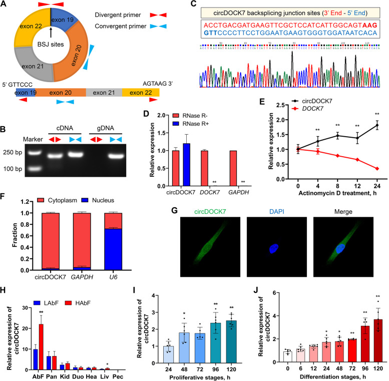
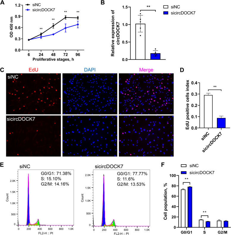
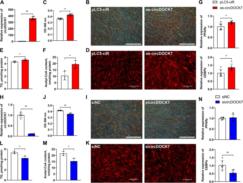
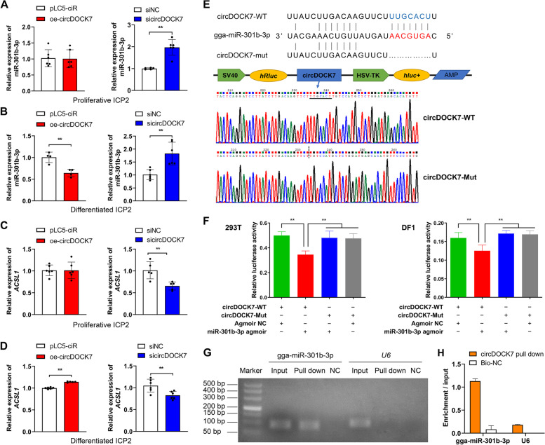
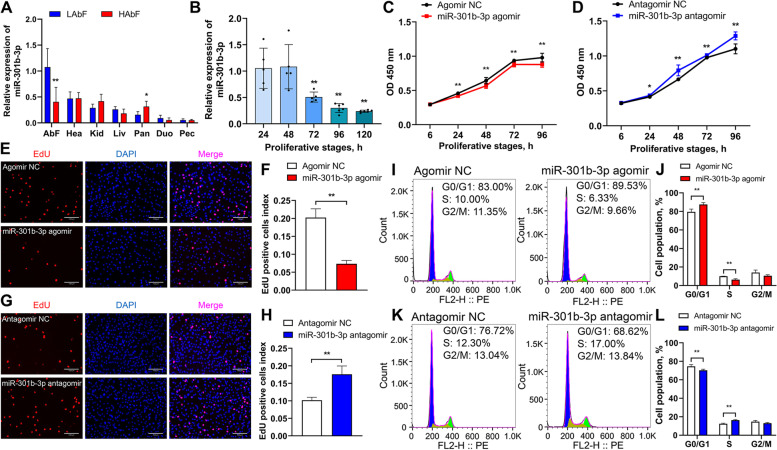
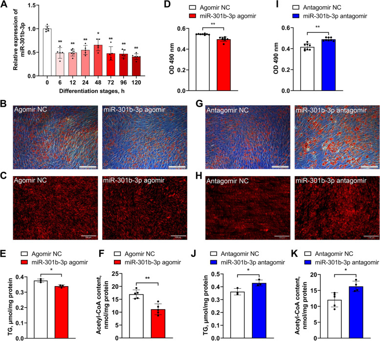
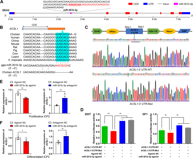
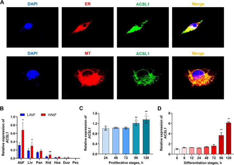
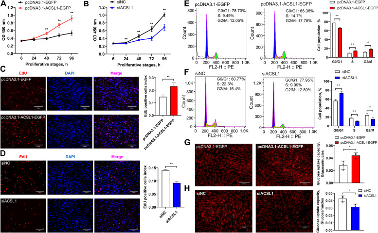
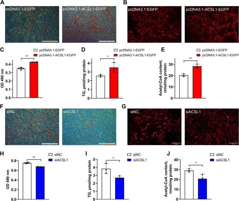
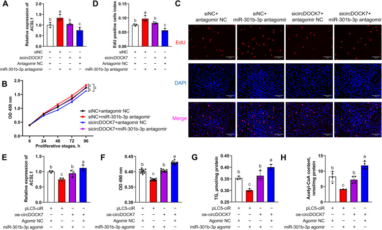
Results: Our results showed that 8:27,886,180|27,889,657 is an exonic circRNA derived from the head-to-tail splicing of exons 19-22 of the dedicator of cytokinesis 7 (DOCK7) gene, abbreviated as circDOCK7. CircDOCK7 is mainly distributed in the cytoplasm of chicken abdominal preadipocytes and is stable because of its RNase R resistance and longer half-life. CircDOCK7 is significantly upregulated in the abdominal fat tissues of fat chickens compared to lean chickens, and its expression gradually increases during the proliferation and adipogenic differentiation of chicken abdominal preadipocytes. Functionally, the gain- and loss-of-function experiments showed that circDOCK7 promoted proliferation, G0/G1- to S-phase progression, and glucose uptake capacity of chicken abdominal preadipocytes, in parallel with adipogenic differentiation characterized by remarkably increased intracellular lipid droplet accumulation and triglyceride and acetyl coenzyme A content in differentiated chicken abdominal preadipocytes. Mechanistically, a pull-down assay and a dual-luciferase reporter assay confirmed that circDOCK7 interacted with gga-miR-301b-3p, which was identified as an inhibitor of chicken abdominal adipogenesis. Moreover, the ACSL1 gene was demonstrated to be a direct target of gga-miR-301b-3p. Chicken ACSL1 protein is localized in the endoplasmic reticulum and mitochondria of chicken abdominal preadipocytes and acts as an adipogenesis accelerator. Rescue experiments showed that circDOCK7 could counteract the inhibitory effects of gga-miR-301b-3p on ACSL1 mRNA abundance as well as the proliferation and adipogenic differentiation of chicken abdominal preadipocytes.

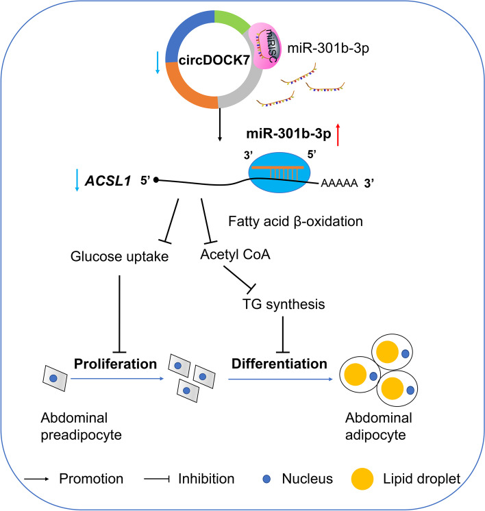
Conclusions: CircDOCK7 serves as a miRNA sponge that directly sequesters gga-miR-301b-3p away from the ACSL1 gene, thus augmenting adipogenesis in chickens. These findings may elucidate a new regulatory mechanism underlying abdominal fat deposition in chickens.

Keywords: Abdominal fat deposition; Adipogenesis; Chickens; CircDOCK7; Competing endogenous RNA; MiRNA sponge.


Conflict of interest statement

The authors declare that they have no competing interests.

Figures

Fig. 1
Fig. 1
Cyclization verification, subcellular localization, and expression pattern analysis of circDOCK7. A Schematic diagram showing the formation and designing of primers for cyclization verification of circDOCK7. B PCR amplification of the divergent and convergent primers of circDOCK7 using gDNA and cDNA as templates, respectively. C Confirmation of the backsplicing junction of circDOCK7 via Sanger’s sequencing. D qRT-PCR analysis of the relative expression of circDOCK7 and DOCK7 in chicken abdominal preadipocytes treated with RNase R. The relative gene expression levels are shown as fold changes compared with that in the RNase R − group. E qRT-PCR analysis of the relative expression of circDOCK7 and DOCK7 in chicken abdominal preadipocytes treated with actinomycin D. The relative gene expression levels are shown as fold changes compared with that in the 0 h group. F qRT-PCR analysis of circDOCK7 expression in the cytoplasm and nuclear fractions of chicken abdominal preadipocytes. G Subcellular localization of circDOCK7 in chicken abdominal preadipocytes using RNA fluorescence in situ hybridization assay. Cell nuclei were stained with 4′,6-diamidino-2-phenylindole DAPI (blue), and circDOCK7 was hybridized with the circDOCK7 probe (green). H Tissue expression pattern of circDOCK7 in chickens with high and low abdominal fat percentage. and J Expression pattern of circDOCK7 during the proliferation and adipogenic differentiation of chicken abdominal preadipocytes. During the proliferation of chicken abdominal preadipocytes, the relative expression levels are shown as fold change versus that of the 24 h group, and the statistical significance analysis are performed versus the 24 h group as control. During the adipogenic differentiation of chicken abdominal preadipocytes, the expression levels are shown as fold change versus that of the 0 h group, and the statistical significance analysis are performed versus the 0 h group as control.  *P < 0.05, **P < 0.01. The same below
Fig. 2
Fig. 2
Effects of circDOCK7 knockdown on the proliferation of chicken abdominal preadipocytes. A CCK8 assay of chicken abdominal preadipocytes transfected with siNC and sicircDOCK7 at 6, 24, 48, 72 and 96 h post-transfection. B Detection of circDOCK7 knockdown in chicken abdominal preadipocytes 48 h post-transfection with sicircDOCK7. The relative gene expression levels are shown as fold changes compared with that in the siNC group. The same below. C Proliferation of chicken abdominal preadipocytes determined by 5-ethynyl-2′-deoxyuridine (EdU) after 48 h of transfection with sicircDOCK7. D Histogram showing the proportion of EdU-positive cells using ImageJ. E and F Cell cycle analysis of chicken abdominal preadipocytes after 48 h transfection with sicircDOCK7 using flow cytometry. *P < 0.05, **P < 0.01
Fig. 3
Fig. 3
Effects of circDOCK7 on the adipogenic differentiation of chicken abdominal preadipocytes. A Detection of circDOCK7 overexpression after 48 h of transfection with circDOCK7 overexpression vector in differentiated chicken abdominal adipocytes. The relative gene expression levels are shown as fold changes compared with that in the pLC5-ciR group. The same below. B and C Representative images of Oil Red O staining and lipid droplet content of circDOCK7-overexpressing differentiated chicken abdominal adipocytes. D Representative images of Nile red fluorescent staining of circDOCK7-overexpressing differentiated chicken abdominal adipocytes. and F Intracellular triglyceride and acetyl-CoA levels of circDOCK7-overexpressing differentiated chicken abdominal adipocytes. G Relative mRNA expression levels of PPARγ and CEBPα in circDOCK7-overexpressing differentiated chicken abdominal adipocytes. H Detection of circDOCK7 knockdown after 48 h of transfection with sicircDOCK7 in differentiated chicken abdominal adipocytes. I and J Representative images of Oil Red O staining and lipid droplet content of circDOCK7-knockdown differentiated chicken abdominal adipocytes. K Representative images of Nile red fluorescent staining of circDOCK7-knockdown differentiated chicken abdominal adipocytes. L and M Intracellular triglyceride and acetyl-CoA levels of circDOCK7-knockdown differentiated chicken abdominal adipocytes. N Relative mRNA expression levels of PPARγ and CEBPα in circDOCK7-knockdown differentiated chicken abdominal adipocytes. *P < 0.05, **P < 0.01
Fig. 4
Fig. 4
circDOCK7 functions as a gga-miR-301b-3p sponge. A and B Relative expression of gga-miR-301b-3p in proliferative and differentiated chicken abdominal preadipocytes upon circDOCK7 overexpression and knockdown. C and D Relative expression of the ACSL1 gene in proliferative and differentiated chicken abdominal preadipocytes upon circDOCK7 overexpression and knockdown. E Construction of dual-luciferase reporter vectors for the validation of gga-miR-301b-3p binding to circDOCK7. WT: wild-type vector; Mut: mutant vector; hRluc: Renilla luciferase; hluc + : firefly luciferase. F Validation of the interaction between gga-miR-301b-3p and circDOCK7 via a dual-luciferase reporter assay in 293 T cells and DF1 cells. G Detection of the combination of circDOCK7 and gga-miR-301b-3p by an RNA pull-down assay. H qRT-PCR analysis of gga-miR-301b-3p expression pulled down by the circDOCK7 probe. *P < 0.05, **P < 0.01
Fig. 5
Fig. 5
Effects of gga-miR-301b-3p on the proliferation of chicken abdominal preadipocytes. A Tissue expression pattern of gga-miR-301b-3p in chickens with high and low abdominal fat percentage. B Expression pattern of gga-miR-301b-3p during the proliferation of chicken abdominal preadipocytes. C CCK8 assay of chicken abdominal preadipocytes transfected with miR-301b-3p agomir and miR-301b-3p agomir NC. D CCK8 assay of chicken abdominal preadipocytes transfected with miR-301b-3p antagomir and miR-301b-3p antagomir NC. E Proliferation of chicken abdominal preadipocytes determined by EdU after 48 h of transfection with miR-301b-3p agomir and miR-301b-3p agomir NC. F Histogram showing the proportion of gga-miR-301b-3p-overexpressing EdU-positive cells using ImageJ. G Proliferation of chicken abdominal preadipocytes determined by EdU after 48 h of transfection with miR-301b-3p antagomir and miR-301b-3p antagomir NC. H Histogram showing the proportion of gga-miR-301b-3p-knockdown EdU-positive cells using ImageJ. I and J Flow-cytometric cell cycle analysis of chicken abdominal preadipocytes after 48 h of transfection with miR-301b-3p agomir and miR-301b-3p agomir NC. K and L Flow-cytometric cell cycle analysis of chicken abdominal preadipocytes after 48 h of transfection with miR-301b-3p antagomir and miR-301b-3p antagomir NC. *P < 0.05, **P < 0.01
Fig. 6
Fig. 6
Effects of gga-miR-301b-3p on the adipogenic differentiation of chicken abdominal preadipocytes. A Expression pattern of gga-miR-301b-3p during the adipogenic differentiation of chicken abdominal preadipocytes. B, C Representative images of Oil Red O staining and Nile red fluorescent staining of gga-miR-301b-3p-overexpressing differentiated chicken abdominal adipocytes. D Detection of lipid droplet content of gga-miR-301b-3p-overexpressing differentiated chicken abdominal adipocytes. E and F Intracellular triglyceride and acetyl-CoA levels of gga-miR-301b-3p-overexpressing differentiated chicken abdominal adipocytes. G and H Representative images of Oil Red O staining and Nile red fluorescent staining of gga-miR-301b-3p-knockdown differentiated chicken abdominal adipocytes. I Detection of the lipid droplet content of gga-miR-301b-3p-knockdown differentiated chicken abdominal adipocytes. and K Intracellular triglyceride and acetyl-CoA levels of gga-miR-301b-3p-knockdown differentiated chicken abdominal adipocytes. *P < 0.05, **P < 0.01
Fig. 7
Fig. 7
Validation of the ACSL1 gene as a direct target of gga-miR-301b-3p. A Schematic diagram of the genomic location of gga-miR-301b-3p. The sequences encoding the precursor gga-miR-301b-3p are shown, with the mature gga-miR-301b-3p sequences highlighted in red. B Complementary sequences of gga-miR-301b-3p and the 3′ UTR of the ACSL1 gene of different species. The seed region of gga-miR-301b-3p is highlighted in red; potential gga-miR-301b-3p-binding sites in the 3′ UTR of the ACSL1 gene are shown in blue. C Construction of dual-luciferase reporter vectors for the validation of gga-miR-301b-3p targeting the ACSL1 gene. WT: wild-type vector; mut: mutant vector; hRluc: Renilla luciferase; hluc + : firefly luciferase. D Validation of the interaction between gga-miR-301b-3p and the 3′ UTR of the ACSL1 gene by a dual-luciferase reporter assay in 293 T cells and DF1 cells. E Relative ACSL1 mRNA expression in gga-miR-301b-3p-overexpressing and -knockdown proliferative chicken abdominal preadipocytes. The relative gene expression levels are shown as fold changes compared with that in the agomir NC and antagomir NC groups, respectively. The same below. F Relative ACSL1 mRNA expression in gga-miR-301b-3p-overexpressing and -knockdown differentiated chicken abdominal preadipocytes. *P < 0.05, **P < 0.01
Fig. 8
Fig. 8
Subcellular localization and expression profiles of chicken ACSL1 protein. A Subcellular localization of ACSL1 in chicken abdominal preadipocytes. MT represents mitochondrion. ER represents endoplasmic reticulum. Cell nuclei were stained with DAPI. B Tissue expression pattern of the ACSL1 gene in chickens with high and low abdominal fat percentage. C and D Expression pattern of ACSL1 during the proliferation and adipogenic differentiation of chicken abdominal preadipocytes. *P < 0.05, **P < 0.01
Fig. 9
Fig. 9
Effects of ACSL1 gene expression on the proliferation of chicken abdominal preadipocytes. A and B CCK8 assay of ACSL1-overexpressing and -knockdown chicken abdominal preadipocytes. and D Proliferation of ACSL1-overexpressing and -knockdown chicken abdominal preadipocytes determined by EdU assay. E and F Flow-cytometric cell cycle analysis of ACSL1-overexpressing and -knockdown chicken abdominal preadipocytes. G and H Glucose uptake capacity of ACSL1-overexpressing and -knockdown chicken abdominal preadipocytes. *P < 0.05, **P < 0.01
Fig. 10
Fig. 10
Effects of ACSL1 gene expression on the adipogenic differentiation of chicken abdominal preadipocytes. A and B Representative images of Oil Red O staining and Nile red fluorescent staining of ACSL1-overexpressing differentiated chicken abdominal adipocytes. C Detection of the lipid droplet content of ACSL1-overexpressing differentiated chicken abdominal adipocytes. D and E Intracellular triglyceride and acetyl-CoA levels of ACSL1-overexpressing differentiated chicken abdominal adipocytes. F and G Representative images of Oil Red O staining and Nile red fluorescent staining of ACSL1-knockdown differentiated chicken abdominal adipocytes. H Detection of the lipid droplet content of ACSL1-knockdown differentiated chicken abdominal adipocytes. I and J Intracellular triglyceride and acetyl-CoA levels of ACSL1-knockdown differentiated chicken abdominal adipocytes. *P < 0.05, **P < 0.01
Fig. 11
Fig. 11
circDOCK7 augments adipogenesis in a gga-miR-301b-3p-dependent manner. A Relative expression of the ACSL1 gene in proliferative chicken abdominal preadipocytes co-transfected with sicircDOCK7 and miR-301b-3p antagomir. The relative gene expression levels are shown as fold changes compared with that in the siNC + antagomir NC group. BD CCK8 and EdU staining assays in proliferative chicken abdominal preadipocytes co-transfected with sicircDOCK7 and miR-301b-3p antagomir. E Relative expression of the ACSL1 gene in differentiated chicken abdominal preadipocytes co-transfected with circDOCK7 overexpression vector and miR-301b-3p agomir. The relative gene expression levels are shown as fold changes compared with that in the pLC5-ciR + agomir NC group. F–H Intracellular lipid droplet accumulation and triglyceride and acetyl-CoA levels in differentiated chicken abdominal adipocytes co-transfected with circDOCK7 overexpression vector and miR-301b-3p agomir. *P < 0.05, **P < 0.01. a–cMeans with different lowercase letters indicate significant difference (P < 0.05)
Fig. 12
Fig. 12
Proposed model for the regulatory mechanism of circDOCK7 in chicken abdominal adipogenesis. CircDOCK7 is a new adipogenesis-stimulating circRNA in chickens. CircDOCK7 serves as a ceRNA to modulate the gga-miR-301b-3p/ACSL1 axis, thus promoting the proliferation and adipogenic differentiation of chicken abdominal preadipocytes

Similar articles

Cited by

References

    1. Zhang XY, Wu MQ, Wang SZ, Zhang H, Du ZQ, Li YM, et al. Genetic selection on abdominal fat content alters the reproductive performance of broilers. Animal. 2018;12(6):1232–1241. doi: 10.1017/S1751731117002658. - DOI - PubMed
    1. Geraert PA, MacLeod MG, Larbier M, Leclercq B. Nitrogen metabolism in genetically fat and lean chickens. Poult Sci. 1990;69(11):1911–1921. doi: 10.3382/ps.0691911. - DOI - PubMed
    1. Demeure O, Duclos MJ, Bacciu N, Le Mignon G, Filangi O, Pitel F, et al. Genome-wide interval mapping using SNPs identifies new QTL for growth, body composition and several physiological variables in an F2 intercross between fat and lean chicken lines. Genet Sel Evol. 2013;45(1):36. doi: 10.1186/1297-9686-45-36. - DOI - PMC - PubMed
    1. Cahaner A, Nitsan Z. Evaluation of simultaneous selection for live body weight and against abdominal fat in broilers. Poult Sci. 1985;64(7):1257–1263. doi: 10.3382/ps.0641257. - DOI - PubMed
    1. Chen LL, Yang L. Regulation of circRNA biogenesis. RNA Biol. 2015;12(4):381–388. doi: 10.1080/15476286.2015.1020271. - DOI - PMC - PubMed

LinkOut - more resources