. 2023 May 10;12(10):1358.
doi: 10.3390/cells12101358.

Mitochondrial Dysfunction and Impaired Antioxidant Responses in Retinal Pigment Epithelial Cells Derived from a Patient with RCBTB1-Associated Retinopathy

Affiliations

Mitochondrial Dysfunction and Impaired Antioxidant Responses in Retinal Pigment Epithelial Cells Derived from a Patient with RCBTB1-Associated Retinopathy

Zhiqin Huang et al. Cells. .

Abstract

Mutations in the RCBTB1 gene cause inherited retinal disease; however, the pathogenic mechanisms associated with RCBTB1 deficiency remain poorly understood. Here, we investigated the effect of RCBTB1 deficiency on mitochondria and oxidative stress responses in induced pluripotent stem cell (iPSC)-derived retinal pigment epithelial (RPE) cells from control subjects and a patient with RCBTB1-associated retinopathy. Oxidative stress was induced with tert-butyl hydroperoxide (tBHP). RPE cells were characterized by immunostaining, transmission electron microscopy (TEM), CellROX assay, MitoTracker assay, quantitative PCR and immunoprecipitation assay. Patient-derived RPE cells displayed abnormal mitochondrial ultrastructure and reduced MitoTracker fluorescence compared with controls. Patient RPE cells displayed increased levels of reactive oxygen species (ROS) and were more sensitive to tBHP-induced ROS generation than control RPE. Control RPE upregulated RCBTB1 and NFE2L2 expression in response to tBHP treatment; however, this response was highly attenuated in patient RPE. RCBTB1 was co-immunoprecipitated from control RPE protein lysates by antibodies for either UBE2E3 or CUL3. Together, these results demonstrate that RCBTB1 deficiency in patient-derived RPE cells is associated with mitochondrial damage, increased oxidative stress and an attenuated oxidative stress response.

Keywords: RCBTB1; inherited retinal disease; mitochondria; oxidative stress; retinal pigment epithelium.


Conflict of interest statement

The authors declare no conflict of interest. The funders had no role in the design of the study; in the collection, analyses, or interpretation of data; in the writing of the manuscript; or in the decision to publish the results.

Figures

Figure 1
Figure 1
Electron microscopic analysis of cultured RPE cells showing morphology and structural features in patient-derived and control RPE cells. (A) A diagram showing the basic structure of RPE cell. (B,C) TEM images of RPE cells demonstrating the ultrastructure of control-(B) and patient-derived (C) RPE. Melanosomes (yellow asterisk) were observed in the apical RPE cytoplasm and nuclei (Nu) in the basal cytoplasm. Apical microvilli, tight junctions, endoplasmic reticulum and abundant mitochondria (blue asterisk) were observed. Note the disrupted cristae formation and architecture of damaged mitochondria (red asterisk) in patient-derived RPE. (D,E) Mean mitochondria length (D) and area (E) were significantly decreased inpatient RPE cells compared with control groups. (F,G) An increase in damaged mitochondria with aberrant cristae was observed in patient RPE cells, compared with control RPE cells. (H,I) Frequency distribution plots of mitochondria by size (H) and area (I). At least 200 mitochondria were measured in each group. Scale bars in (B,C,G) indicate 1 µm. *** p < 0.0001.
Figure 2
Figure 2
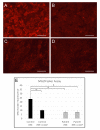
MitoTracker labelling of RPE mitochondria. (AD) Fluorescence micrographs showing live control- (A,B) and patient-derived (C,D) RPE monolayers labelled with MitoTracker Orange CMTMRos reagent. Panels on the right (B,D) were treated with 10 μM CCCP. Scale bars indicate 100 μm. (E) Bar graph showing mean MitoTracker fluorescence intensities in control- and patient-derived RPE cells, with and without CCCP treatment. Each bar represents the mean Mitotracker signal from RPE monolayers derived from three independent iPSC lines. Error bars indicate standard deviation. * p < 0.05.
Figure 3
Figure 3
Response of RPE cells to oxidative stress induced by tBHP. Control- and patient-derived RPE monolayers were treated with 0, 100 or 200 μM of tBHP for 1 h. (A) ROS levels in live control- and patient-derived RPE monolayers were measured by CellROX assay. (B–F) Expression of RCBTB1 (B), NFE2L2 (C), IDH1 (D), SLC25A25 (E) and RXRA (F) was measured by qPCR. Each bar represents the mean values obtained from RPE cells generated from three independently derived iPSC lines. Error bars indicate standard deviation. * p < 0.05; ** p < 0.01.
Figure 4
Figure 4
Western blots showing immunoprecipitation results from control iPSC-derived RPE. Protein lysates from control RPE cultures were immunoprecipitated using beads conjugated with either anti-CUL3 (left side) or anti-UBE2E3 (right side) antibodies and analysed by Western blotting using anti-CUL3 (top), anti-RCBTB1 (middle) or anti-UBE2E3 (bottom). Western blot analysis demonstrated coimmunoprecipitation of cullin-3, UBE2E3 and RCBTB1 proteins in RPE cells derived from healthy control iPSC lines using both antibodies. Lanes: M: Protein ladder; I: 10 μg input protein; N: control immunoprecipitation with beads alone (no conjugated antibody, elution 2); E1: Immunoprecipitation with antibody-conjugated beads, elution 1 (without beta-mercaptoethanol); E2: Immunoprecipitation with antibody-conjugated beads, elution 2 (with beta-mercaptoethanol).

References

    1. Wu J.H., Liu J.H., Ko Y.C., Wang C.T., Chung Y.C., Chu K.C., Liu T.T., Chao H.M., Jiang Y.J., Chen S.J., et al. Haploinsufficiency of RCBTB1 is associated with Coats disease and familial exudative vitreoretinopathy. Hum. Mol. Genet. 2016;25:1637–1647. doi: 10.1093/hmg/ddw041. - DOI - PubMed
    1. Coppieters F., Ascari G., Dannhausen K., Nikopoulos K., Peelman F., Karlstetter M., Xu M., Brachet C., Meunier I., Tsilimbaris M.K., et al. Isolated and Syndromic Retinal Dystrophy Caused by Biallelic Mutations in RCBTB1, a Gene Implicated in Ubiquitination. Am. J. Hum. Genet. 2016;99:470–480. doi: 10.1016/j.ajhg.2016.06.017. - DOI - PMC - PubMed
    1. Huang Z., Zhang D., Thompson J.A., Jamuar S.S., Roshandel D., Jennings L., Mellough C., Charng J., Chen S.C., McLaren T.L., et al. Deep clinical phenotyping and gene expression analysis in a patient with RCBTB1-associated retinopathy. Ophthalmic Genet. 2021;42:266–275. doi: 10.1080/13816810.2021.1891551. - DOI - PubMed
    1. Yang J., Xiao X., Sun W., Li S., Jia X., Zhang Q. Variants in RCBTB1 are Associated with Autosomal Recessive Retinitis Pigmentosa but Not Autosomal Dominant FEVR. Curr. Eye Res. 2020;46:839–844. doi: 10.1080/02713683.2020.1842457. - DOI - PubMed
    1. Catomeris A.J., Ballios B.G., Sangermano R., Wagner N.E., Comander J.I., Pierce E.A., Place E.M., Bujakowska K.M., Huckfeldt R.M. Novel RCBTB1 variants causing later-onset non-syndromic retinal dystrophy with macular chorioretinal atrophy. Ophthalmic Genet. 2022;43:332–339. doi: 10.1080/13816810.2021.2023196. - DOI - PMC - PubMed

Publication types

MeSH terms