. 2023 May 13;12(10):1380.
doi: 10.3390/cells12101380.

Temporal Changes in Nuclear Envelope Permeability during Semi-Closed Mitosis in Dictyostelium Amoebae

Affiliations

Temporal Changes in Nuclear Envelope Permeability during Semi-Closed Mitosis in Dictyostelium Amoebae

Kristina Mitic et al. Cells. .

Abstract

The Amoebozoan Dictyostelium discoideum exhibits a semi-closed mitosis in which the nuclear membranes remain intact but become permeabilized to allow tubulin and spindle assembly factors to access the nuclear interior. Previous work indicated that this is accomplished at least by partial disassembly of nuclear pore complexes (NPCs). Further contributions by the insertion process of the duplicating, formerly cytosolic, centrosome into the nuclear envelope and nuclear envelope fenestrations forming around the central spindle during karyokinesis were discussed. We studied the behavior of several Dictyostelium nuclear envelope, centrosomal, and nuclear pore complex (NPC) components tagged with fluorescence markers together with a nuclear permeabilization marker (NLS-TdTomato) by live-cell imaging. We could show that permeabilization of the nuclear envelope during mitosis occurs in synchrony with centrosome insertion into the nuclear envelope and partial disassembly of nuclear pore complexes. Furthermore, centrosome duplication takes place after its insertion into the nuclear envelope and after initiation of permeabilization. Restoration of nuclear envelope integrity usually occurs long after re-assembly of NPCs and cytokinesis has taken place and is accompanied by a concentration of endosomal sorting complex required for transport (ESCRT) components at both sites of nuclear envelope fenestration (centrosome and central spindle).

Keywords: Dictyostelium; centrosome; mitosis; nuclear envelope; nuclear pore complex.


Conflict of interest statement

The authors declare no conflict of interest.

Figures

Figure 1
Figure 1
NLS-TdTom is only inside the nucleus during interphase. Selected time points of Video S2. NLS-TdTom (magenta) diffuses over the whole cell upon permeabilization of the nucleus at the onset of mitosis (60 s, arrow). Cells co-expressing GFP-α-tubulin (green) are observed to show the mitotic progression. The nuclear marker is diffused completely before the centrosome duplication (arrowhead) becomes visible (310 s). After cytokinesis, NLS-TdTom is located back to the nucleus (1110 s). Bar 5 µm.
Figure 2
Figure 2
Nuclear envelope permeabilization and centrosome duplication occur at the onset of mitosis. Selected time points of Videos S3 and S4 are shown. (A) NE marker Src1-Neon knock-in (green) with the NE permeabilization marker NLS-TdTom (magenta). The nuclear envelope is preserved during the entire cell cycle, while NLS-TdTom diffuses into the cytosol (arrow at 100 s) and is restored long after cytokinesis (920 s). (B) Cell with Src1-Neon knock-in (green) and centrosomal component CP75-Ruby knock-in (magenta). The centrosome moves from a perinuclear position (−67.2 s) into the nuclear envelope (−33.6 s) before it duplicates (arrowhead at 0 s). Bars 5 µm.
Figure 3
Figure 3
NLS-TdTom diffuses into the cytosol prior to centrosome splitting. (A) Selected time points from live-cell imaging of a strain expressing CP75-GFP (green) knock-in together with the NE permeabilization marker NLS-TdTom (magenta). NLS-TdTom is located within the nucleus during interphase (−30 s) and diffuses rapidly into the cytosol upon permeabilization (arrow at −20 s and 0 s) while CP75-GFP is still located at the mitotic centrosome only until the splitting of the core structures at time point 0 s (indicated by zoomed inset). Series of frames based on confocal spinning disk live-cell microscopy were selected from a movie (z-distance of 0.5 µm, recorded with a time lapse of 10 s). Only 1 slice out of a 10-layer Z-stack is shown, respectively. Bar 5 µm. See also Video S7. (B) Time resolution between the first NE permeabilization and the duplication of the mitotic centrosome (time1) and the corresponding time interval of image acquisition (time2). Centrosome duplication starts approximately 39 ± 7.3 s (mean and standard deviation for n = 4) after permeabilization of the nuclear envelope.
Figure 4
Figure 4
Nup62-Scarlet disassembly from NPCs precedes centrosome splitting at the onset of mitosis. Selected time points from a live-cell imaging (Video S8) show a cell co-expressing the Nup62-Scarlet (magenta) and CP75-GFP (green) knock-in construct. CP75-GFP is still located at the mitotic centrosome when Nup62-Scarlet starts to dissociate from the NPCs (−30 s, arrow). The time point of 0 s indicates the centrosome splitting which appears as 2 dots (arrowhead). Nup62 dissociation occurred approx. 33 ± 8.4 s (mean and standard deviation for n = 9) prior to centrosome duplication (see Table S3). CP75-GFP leaves the centrosome after the duplication process (40 s). Image stacks of 10 layers (z-distance of 0.5 µm) were recorded with a 10 s time lapse and only 1 focal plane is presented. Bar 5 µm.
Figure 5
Figure 5
NE permeabilization versus Nup62 disassembly during mitosis. (A) Selected time points from live-cell imaging (Video S10) of a strain co-expressing the Nup62-Neon knock-in construct (green) and the NE permeabilization marker NLS-TdTom (magenta). NLS-TdTom leaves the nucleus at the onset of mitosis while Nup62-Neon is still present at the NE (arrow at 20 s). Nup62-Neon completely disassembles from the NE (arrow at 80 s) before it reappears in metaphase (arrow at 410 s). Out of a 12-layer image stack (z-distance 0.5 µm), only the focus plane is presented, respectively. Images were recorded with a 10 s time lapse. Bar 5 µm. (B) The graph presents the fluorescence intensity values for Nup62-Neon knock-in and NLS-TdTom over the complete cell division in (A).
Figure 6
Figure 6
ESCRT proteins concentrate at the nuclear envelope fenestration sites in mitosis and long after cytokinesis has taken place. (A) Cells were fixed with glutaraldehyde and stained with Hoechst (blue) and anti-α-tubulin (magenta). ESCRT-Neon fusion proteins (green) localize at the nuclear envelope fenestration sites in telophase (arrows). A maximum intensity projection of several slices of the deconvolved Z-stack is presented. (B) Selected time points of Video S13 are shown with cells in cytokinesis (−154 s), after cell division (0 s), and once AlxA-Neon has disappeared together with reappearance of NLS-TdTom (arrowhead) at 363 s. Bars 5 µm.
Figure 7
Figure 7
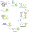
Hypothesis of centrosome insertion and nuclear envelope permeabilization in Dictyostelium discoideum. The onset of mitosis is characterized by the loss of the corona together with the attached microtubules in early prophase. The fusion of the inner and outer nuclear membranes forms a fenestra and allows the insertion of the mitotic centrosome into the nuclear envelope. Simultaneously, the partial disassembly of the nuclear pore complexes contributes to the permeabilization of the nuclear envelope in late prophase. Next, in prometaphase, the central layer of the core structure disappears, the mitotic centrosome splits, and the outer entities move apart. During telophase, the central core layer re-appears and the nuclear pore complexes are reassembled. The duplicated centrosomes exit their fenestrae in the nuclear envelope and the ESCRT-III complex starts re-sealing the nuclear envelope to close the fenestrae.

Similar articles

Cited by

References

    1. Sazer S., Lynch M., Needleman D. Deciphering the Evolutionary History of Open and Closed Mitosis. Curr. Biol. 2014;24:R1099-103. doi: 10.1016/j.cub.2014.10.011. - DOI - PMC - PubMed
    1. Adl S.M., Bass D., Lane C.E., Lukeš J., Schoch C.L., Smirnov A., Agatha S., Berney C., Brown M.W., Burki F., et al. Revisions to the Classification, Nomenclature, and Diversity of Eukaryotes. J. Eukaryot. Microbiol. 2019;66:4–119. doi: 10.1111/jeu.12691. - DOI - PMC - PubMed
    1. Stafstrom J.P., Staehelin L.A. Dynamics of the Nuclear Envelope and of Nuclear Pore Complexes during Mitosis in the Drosophila Embryo. Eur. J. Cell Biol. 1984;34:179–189. - PubMed
    1. De Souza C.P., Osmani A.H., Hashmi S.B., Osmani S.A. Partial Nuclear Pore Complex Disassembly during Closed Mitosis in Aspergillus Nidulans. Curr. Biol. 2004;14:1973–1984. doi: 10.1016/j.cub.2004.10.050. - DOI - PubMed
    1. Mitic K., Grafe M., Batsios P., Meyer I. Partial Disassembly of the Nuclear Pore Complex Proteins during Semi-Closed Mitosis in Dictyostelium Discoideum. Cells. 2022;11:407. doi: 10.3390/cells11030407. - DOI - PMC - PubMed

Publication types

LinkOut - more resources