. 2023 May 13;12(10):1383.
doi: 10.3390/cells12101383.

Co-Stimulation of AGEs and LPS Induces Inflammatory Mediators through PLCγ1/JNK/NF-κB Pathway in MC3T3-E1 Cells

Affiliations

Co-Stimulation of AGEs and LPS Induces Inflammatory Mediators through PLCγ1/JNK/NF-κB Pathway in MC3T3-E1 Cells

Natsuko Tanabe et al. Cells. .

Abstract

Advanced glycation end-products (AGEs) are increased under hyperglycemia in vivo and are associated with the onset of diabetes. According to previous studies, AGEs exacerbate inflammatory diseases. However, the mechanism by which AGEs aggravate osteoblast inflammation remains unknown. Therefore, the aim of this study was to determine the effects of AGEs on the production of inflammatory mediators in MC3T3-E1 cells and the underlying molecular mechanisms. Co-stimulation with AGEs and lipopolysaccharide (LPS) was found to increase the mRNA and protein levels of cyclooxygenase 2 (COX2), interleukin-1α (IL-1α), S100 calcium-binding protein A9 (S100A9), and the production of prostaglandin E2 (PGE2) compared to no stimulation (untreated control) or individual stimulation with LPS or AGEs. In contrast, the phospholipase C (PLC) inhibitor, U73122, inhibited these stimulatory effects. Co-stimulation with AGEs and LPS also increased the nuclear translocation of nuclear factor-kappa B (NF-κB) compared to no stimulation (untreated control) or individual stimulation with LPS or AGE. However, this increase was inhibited by U73122. Co-stimulation with AGEs and LPS-induced phosphorylated phospholipase Cγ1 (p-PLCγ1) and phosphorylated c-Jun N-terminal kinase (p-JNK) expression compared to no stimulation or individual stimulation with LPS or AGEs. U73122 inhibited the effects induced by co-stimulation. siPLCγ1 did not increase the expression of p-JNK and the translocation of NF-κB. Overall, co-stimulation with AGEs and LPS may promote inflammation mediators in MC3T3-E1 cells by activating the nuclear translocation of NF-κB via PLCγ1-JNK activation.

Keywords: AGEs; IL-1α; JNK; LPS; NF-κB; PGE2; PLCγ1; S100A9.


Conflict of interest statement

The authors declare no conflict of interest. The funders had no role in the design of the study; in the collection, analyses, or interpretation of data; in the writing of the manuscript; or in the decision to publish the results.

Figures
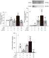
Figure 1
Figure 1
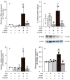
Cells stimulated with or without AGEs (100 μg/mL) and LPS (100 ng/mL), treated with U73122 (10 μM), or not stimulated (untreated control). The mRNA expression of COX2 (A) on day 14 of culture based on real-time PCR. The protein expression of COX2 (B) on day 14 of culture based on western blotting. The production of PGE2 (C) on day 14 based on ELISA. Data are expressed as the mean ± SD of three independent experiments performed in triplicate. One-way ANOVA was employed for comparisons between groups while Tukey’s post hoc test was employed for multiple comparisons among all groups. * p < 0.05, ** p < 0.01 vs. untreated, ## p < 0.01, ### p < 0.001 vs. LPS, ++ p < 0.01 vs. AGEs,  p < 0.05, ★★★ p < 0.001 vs. LPS+AGEs.
Figure 2
Figure 2
Cells stimulated with or without AGEs (100 μg/mL) and LPS (100 ng/mL), treated with U73122 (10 μM), or not stimulated (untreated control). The mRNA expression of IL-1α (A) and S100A9 (C) on day 14 of culture based on real-time PCR. The production of IL-1α (B) on day 14 of culture based on ELISA. The protein expression of S100A9 (D) on day 14 based on western blotting. Data are expressed as the mean ± SD of three or four independent experiments performed in triplicate. One-way ANOVA was employed for comparisons between groups while Tukey’s post hoc test was employed for multiple comparisons among all groups. * p < 0.05, ** p < 0.01, *** p < 0.001 vs. untreated, # p < 0.05, ## p < 0.01 vs. LPS, + p < 0.05, ++ p < 0.01 +++ p < 0.001 vs. AGEs,  p < 0.05, ★★ p < 0.01, ★★★ p < 0.001 vs. LPS+AGEs.
Figure 3
Figure 3
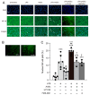
Cells plated on coverslips and stimulated with or without AGEs (100 μg/mL) and LPS (100 ng/mL), treated with U73122 (10 μM) or FPS-ZM1 (40 μM), or not stimulated (untreated control) for 30 min, were fixed with methanol. NF-κB localization was assessed using immunofluorescence. Image of LPS+AGEs-stimulated cells; arrows indicate that NF-κB localized in the nuclei (A). Image of a osteoblasts cytoplasmic (cyto) NF-κB localization. Images of osteoblasts showing NF-κB localized in the nuclei (nuclei) (B). Bar graph showing the percentage of cells with nuclear localization of NF-κB (C). Data are expressed as the mean ± SD of three independent experiments performed in triplicate. One-way ANOVA was employed for comparisons between groups while Tukey’s post hoc test was employed for multiple comparisons among all groups. *** p < 0.001 vs. untreated, ## p < 0.01 vs. LPS, + p < 0.05, +++ p < 0.001 vs. AGEs,  p < 0.05 vs. LPS+AGEs.
Figure 4
Figure 4
Cells stimulated with or without AGEs (100 μg/mL) and LPS (100 ng/mL), and treated with U73122 (10 μM), or not stimulated (untreated control) for 10 and 15 min. The protein expression of p-PLCγ1 (A) and p-JNK (B) based on western blotting. Data are expressed as the mean ± SD of three or four independent experiments performed in triplicate. One-way ANOVA was employed for comparisons between groups while Tukey’s post hoc test was employed for multiple comparisons among all groups. * p < 0.05 vs. untreated, # p < 0.05 vs. LPS, + p < 0.05 vs. AGEs.
Figure 5
Figure 5
Cells were transfected with PLCγ1-specific siRNA (siPLCγ1) or control scrambled siRNA (siControl). The expression of PLCγ1 mRNA and protein based on real-time PCR (A) and western blotting (B). Cells stimulated with AGEs (100 μg/mL) and LPS (100 ng/mL) or not stimulated (untreated control) for 10 and 15 min. The protein expression of p-JNK (C) based on western blotting. Data are expressed as the mean ± SD of three independent experiments performed in triplicate. Unpaired t-test was employed for comparisons between two groups. * p < 0.05, *** p < 0.001 vs. siControl untreated, # p < 0.05, ### p < 0.001 vs. siControl LPS, +++ p < 0.001 vs. siControl LPS+AGEs.
Figure 6
Figure 6
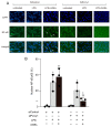
Cells were transfected with PLCγ1-specific siRNA (siPLCγ1) or control scrambled siRNA (siControl) and either stimulated or not stimulated with AGEs (100 μg/mL) and LPS (10 μg/mL). After 30 min of AGEs and LPS co-stimulation, the cells were fixed with methanol. NF-κB localization was assessed using immunofluorescence. Image of LPS+AGEs-stimulated cells; arrows indicate that NF-κB localized in the nuclei (A). Bar graph showing the percentage of cells exhibiting nuclear localization of NF-κB (B). Data are expressed as the mean ± SD of three independent experiments performed in triplicate. One-way ANOVA was employed for comparisons between groups while Tukey’s post hoc test was employed for multiple comparisons among all groups. * p < 0.05, ** p < 0.01 vs. siControl untreated, # p < 0.05 vs. siControl LPS, ++ p < 0.01 vs. siControl LPS+AGEs, p < 0.05 vs. siPLCγ1 LPS.

Similar articles

Cited by

References

    1. Twarda-clapa A., Olczak A., Białkowska A.M., Koziołkiewicz M. Advanced Glycation End-Products (AGEs): Formation, Chemistry, Classification, Receptors, and Diseases Related to AGEs. Cells. 2022;11:1312. doi: 10.3390/cells11081312. - DOI - PMC - PubMed
    1. Khalid M., Petroianu G., Adem A. Advanced Glycation End Products and Diabetes Mellitus: Mechanisms and Perspectives. Biomolecules. 2022;12:542. doi: 10.3390/biom12040542. - DOI - PMC - PubMed
    1. Sato T., Iwaki M., Shimogaito N., Wu X., Yamagishi S., Takeuchi M. TAGE (Toxic AGEs) Theory in Diabetic Complications. Curr. Mol. Med. 2006;6:351–358. doi: 10.2174/156652406776894536. - DOI - PubMed
    1. Miura J., Yamagishi S., Uchigata Y., Takeichi M., Yamamoto H., Makita Z., Iwamoto Y. Serum Levels of Non-carboxymethyllysine Advanced Glycation Endproducts are Correlated to Severity of Microvascular Complications in Patients with Type 1 Diabetes. J. Diabetes Complicat. 2003;17:16–21. doi: 10.1016/S1056-8727(02)00183-6. - DOI - PubMed
    1. Dariya B., Nagaraju G.P. Advanced Glycation End Products in Diabetes, Cancer and Phytochemical Therapy. Drug Discov. Today. 2020;25:1614–1623. doi: 10.1016/j.drudis.2020.07.003. - DOI - PubMed

Publication types

MeSH terms