

Published Erratum
doi: 10.3390/cells12101356.
Correction: Garza-Lopez et al. Protocols for Generating Surfaces and Measuring 3D Organelle Morphology Using Amira. Cells 2022, 11, 65
Affiliations
- PMID: 37408281
- PMCID: PMC10216418
- DOI: 10.3390/cells12101356
Item in Clipboard
Published Erratum
Correction: Garza-Lopez et al. Protocols for Generating Surfaces and Measuring 3D Organelle Morphology Using Amira. Cells 2022, 11, 65
Cells.
.
Abstract
In the original publication [...].
Figures
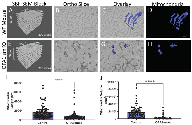
Skeletal muscle specific knockout of OPA1 (OPA1 smKO) in mouse leads to changes in mitochondrial morphology in the mouse. The 3D distribution of single continuous and stationary mitochondria (blue), reconstructed from serial block facing-scanning electron microscopy (SBF-SEM) image stacks of gastrocnemius muscle from OPA1 smKO mouse (A–H). (A) The dimensions of the captured tissue in wild type mouse and (E) OPA1 smKO, (B,F) along with an example ortho slice for each. (C) The overlay of the 3D surface rendering of mitochondria in a wild type mouse, on top of a representative ortho slice and (D) the 3D surface rendering of mitochondria alone. (G) The overlay of the 3D rendering of mitochondria in OPA1 smKO, on top of a representative ortho slice and (H) the 3D surface rendering of mitochondria alone. (I,J) The 3D mitochondrial length and volume decreased (**** p < 0.001) upon OPA1 smKO.
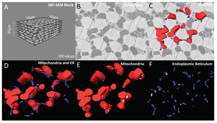
6-panel presentation of 3D reconstruction images and ortho slices from wildtype Drosophila flight muscle. This figure is an example of how to present the ortho slices and the 3D reconstruction images. This example shows 3D reconstruction of several organelles in Drosophila flight muscle. (A) On the left, several representative ortho slices are presented. The dimensions and amounts of ortho slices for data acquisition and conversion to 3D models are shown. (B) The raw image of an ortho slice. (C–F) Mitochondria are colored red, ER are colored blue, and MERCs are colored white. These data are best presented in several ways. (C) 3D reconstruction overlaid over the ortho image allows for better visualization of the specific structures in the ortho image that are reconstructed. (D) In contrast, the 3D reconstruction not overlaid on the ortho image allows for better visualization of interactions between the 3D structures. (E,F) Finally, Amira also allows for the graying out of specific structures such that only mitochondria or ER are shown in the 3D reconstruction. This is useful to view otherwise difficult to see areas including MERCs.
Erratum for
-
Protocols for Generating Surfaces and Measuring 3D Organelle Morphology Using Amira.Cells. 2021 Dec 27;11(1):65. doi: 10.3390/cells11010065. Cells. 2021. PMID: 35011629 Free PMC article.
References
Publication types
Grants and funding
LinkOut - more resources
Full Text Sources
