

Kaposiform hemangioendothelioma of the thigh: A case report
- PMID: 37409100
- PMCID: PMC10318450
- DOI: 10.1016/j.radcr.2023.05.052
Kaposiform hemangioendothelioma of the thigh: A case report
Abstract
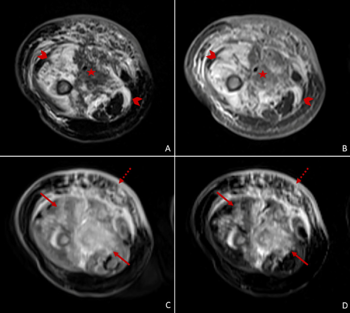
Kaposiform hemangioendothelioma is a rare, locally aggressive or borderline vascular tumor that typically affects infants. It presents as a purpuric cutaneous lesion and may be associated with life-threatening coagulation disorders, such as the Kasabach-Merritt phenomenon. The differential diagnosis can be challenging based on clinical presentation alone. Imaging plays a crucial role in the diagnostic workup, particularly magnetic resonance imaging. We present a case report of a 4-month-old patient with an enlarging vinous cutaneous mass on the thigh and coagulation abnormalities. Magnetic resonance imaging revealed a large, infiltrative, soft-tissue lesion with poorly defined margins and heterogeneous enhancement, that involved all muscle compartments of the thigh and was associated with lymphedema, stranding of the subcutaneous fat and cutaneous thickening. These findings were consistent with kaposiform hemangioendothelioma of the thigh and the diagnosis was confirmed by histopathological characterization.
Keywords: Kaposiform hemangioendothelioma; Kasabach-Merritt Syndrome; Magnetic resonance imaging.
© 2023 The Authors. Published by Elsevier Inc. on behalf of University of Washington.
Figures


Similar articles
-
Neonatal kaposiform hemangioendothelioma of the spleen associated with Kasabach-Merritt phenomenon.J Pediatr Surg. 2016 Jun;51(6):1047-50. doi: 10.1016/j.jpedsurg.2016.03.014. Epub 2016 Apr 9. J Pediatr Surg. 2016. PMID: 27342010
-
Multidisciplinary management of a neonate with kaposiform hemangioendothelioma with extensive cranial fossa destruction.SAGE Open Med Case Rep. 2022 Dec 17;10:2050313X221142685. doi: 10.1177/2050313X221142685. eCollection 2022. SAGE Open Med Case Rep. 2022. PMID: 36545011 Free PMC article.
-
Imaging findings of Kaposiform Hemangioendothelioma in children.Eur J Radiol. 2017 Jan;86:198-205. doi: 10.1016/j.ejrad.2016.11.015. Epub 2016 Nov 10. Eur J Radiol. 2017. PMID: 28027747
-
Multivertebral Kaposiform hemangioendothelioma presenting as scoliosis - A case report and review of literature.J Clin Orthop Trauma. 2023 Mar 17;39:102147. doi: 10.1016/j.jcot.2023.102147. eCollection 2023 Apr. J Clin Orthop Trauma. 2023. PMID: 37021123 Free PMC article. Review.
-
Intracranial kaposiform hemangioendothelioma presenting as epistaxis: a rare case report with review of literature.Childs Nerv Syst. 2021 Jun;37(6):2057-2062. doi: 10.1007/s00381-020-04905-y. Epub 2020 Sep 28. Childs Nerv Syst. 2021. PMID: 32989498 Review.
References
-
- International Society for the Study of Vascular Anomalies ISSVA classification of vascular anomalies. issva.org/classification; 2018 [accessed 15.09.23].
Publication types
LinkOut - more resources
Full Text Sources
