. 2023 Jun 20:17:1167007.
doi: 10.3389/fnins.2023.1167007. eCollection 2023.

Investigation of the efficacy and safety of retinal inactivation as a treatment for amblyopia in cats

Affiliations

Investigation of the efficacy and safety of retinal inactivation as a treatment for amblyopia in cats

Mairin Hogan et al. Front Neurosci. .

Abstract

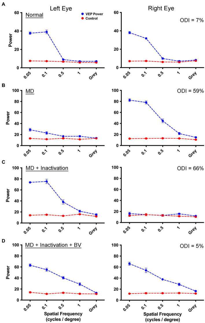
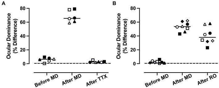
Introduction: Deprivation of normal vision early in postnatal development elicits modifications of neural circuitry within the primary visual pathway that can cause a severe and intractable vision impairment (amblyopia). In cats, amblyopia is often modeled with monocular deprivation (MD), a procedure that involves temporarily closing the lids of one eye. Following long-term MD, brief inactivation of the dominant eye's retina can promote recovery from the anatomical and physiological effects of MD. In consideration of retinal inactivation as a viable treatment for amblyopia it is imperative to compare its efficacy against conventional therapy, as well as assess the safety of its administration.

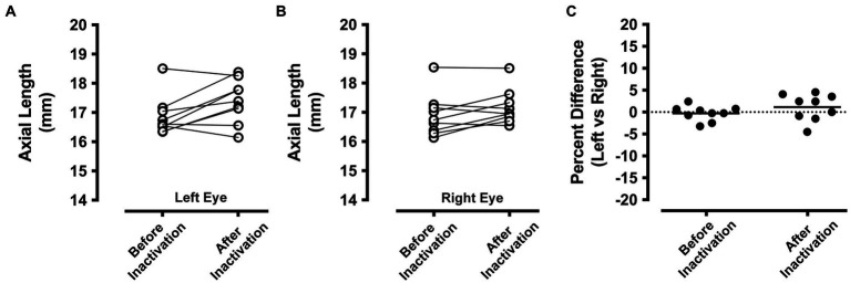
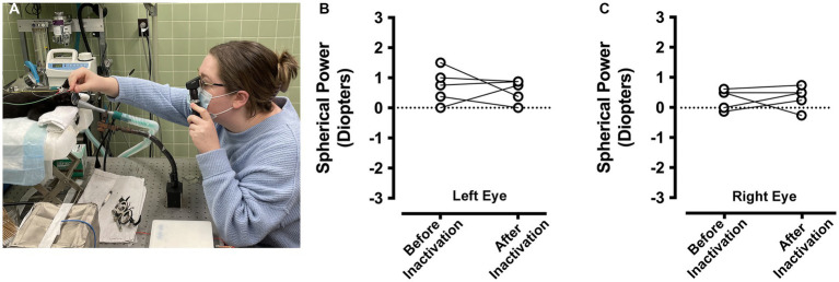
Methods: In the current study we compared the respective efficacies of retinal inactivation and occlusion of the dominant eye (reverse occlusion) to elicit physiological recovery from a prior long-term MD in cats. Because deprivation of form vision has been associated with development of myopia, we also examined whether ocular axial length or refractive error were altered by a period of retinal inactivation.

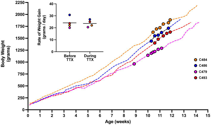
Results: The results of this study demonstrate that after a period of MD, inactivation of the dominant eye for up to 10 days elicited significant recovery of visually-evoked potentials that was superior to the recovery measured after a comparable duration of reverse occlusion. After monocular retinal inactivation, measurements of ocular axial length and refractive error were not significantly altered from their pre-inactivation values. The rate of body weight gain also was not changed during the period of inactivation, indicating that general well-being was not affected.

Discussion: These results provide evidence that inactivation of the dominant eye after a period of amblyogenic rearing promotes better recovery than eye occlusion, and this recovery was achieved without development of form-deprivation myopia.

Keywords: amblyopia; ocular axial length; plasticity; refractive error; retinal inactivation; tetrodotoxin; visual cortex; visually-evoked potentials.


Conflict of interest statement

The authors declare that the research conducted for this study has no commercial or financial relationships that could be viewed as a conflict of interest.

Figures

Figure 1
Figure 1
Measurement of VEPs from the left V1 elicited by separate stimulation of the left and right eye in a monocularly deprived animal that received fellow eye inactivation. For each graph, spatial frequency is plotted on the abscissa, and the summed power from the Fourier analysis is plotted on the ordinate. The blue trace represents the sum of visually-evoked power, while the red trace shows the non-visual baseline power. Visually-evoked power elicited by a gray screen served as a control, and should be about equal for the blue and red traces. Data are shown for an example animal (C479) in which VEP power from the left and right eye were balanced (blue traces) prior to any visual manipulation (A). Following 3 weeks of left eye MD, there was a strong shift in ocular dominance with VEPs obviously attenuated for the deprived eye, and potentiated for the fellow non-deprived eye (B). Inactivation of the non-deprived (right) eye for 8 days produced a recovery of VEPs in the originally deprived eye, while VEPs serving the inactivated eye remained at baseline levels due to the continued effect of TTX (C). Following a washout period of 2 weeks, VEPs serving the inactivated eye were restored and in balance with those from the originally deprived eye (D). For all animals in this study, VEPs exhibited lower power at the youngest age examined (A) compared to older animals (D). It is unclear what produced this difference but it could be the result of a progressive maturation of V1 from 4 weeks of age to about 12 weeks of age. The percentage difference between VEPs measured between the two eyes across all spatial frequencies (ODI) for each rearing condition is displayed in the upper right corner of the right eye VEP graphs.
Figure 2
Figure 2
Measurement of VEPs from left V1 elicited by separate stimulation of the left and right eye in a monocularly deprived animal that received RO. Symbol and graph details are the same as those for Figure 1. Data are shown for an example animal (C481) in which VEP power from the left and right eye are balanced prior to any visual manipulation (A). VEPs measured after 3 weeks of left-eye MD were attenuated for the deprived eye, but were potentiated for the fellow non-deprived eye indicative of a substantial shift in ocular dominance (B). VEPs measured from the originally deprived eye did not increase appreciably after RO (C), and measurements from originally non-deprived eye were attenuated to baseline because recordings were made while the eye was still closed. After the eye was opened and binocular vision was provided for 4 days, VEPs measured from the originally deprived eye remained diminished, while VEPs elicited from the eye that received RO were restored to their potentiated state before the RO was imposed (D). This indicated that 10 days of RO imposed after 3 weeks of MD was insufficient to produce a substantial recovery of the deprived eye. The percentage difference between VEPs measured between the two eyes (ODI) for each rearing condition is displayed in the upper right corner of the right eye VEP graphs.
Figure 3
Figure 3
Comparison of the recovery promoted by MI or RO following 3 weeks of MD. Recovery was assessed with a calculation of ocular dominance that involved calculating the percentage difference in summed VEP power across all spatial frequencies that were presented separately to the left and right eye (see the section Materials and methods). For the MI group (n = 3 animals), balanced ocular dominance measured before MD was altered significantly after 3 weeks because VEP power was reduced by 68% in the deprived eye relative to the non-deprived eye (A). After 3 weeks of MD, animals that had their fellow eye inactivated showed a recovery of normal ocular dominance in which summed VEP power was not different between the eyes. The comparison group of RO animals (n = 4) also showed a shift in ocular dominance so that VEP power for the deprived eye was reduced by 54%. Ten days of RO after the period of MD did not restore normal ocular dominance (B). Open and solid symbols represent left and right V1, respectively. Symbol shapes represent different animals.
Figure 4
Figure 4
Ocular axial length measurements before and after inactivation of the right eye (n = 9 animals). A-scan measurements of the left eye that were obtained before MI were comparable to those taken after the period of inactivation (A). A similar result was observed for the right eye, which showed before inactivation axial length measurements that were similar to those taken after inactivation (B). Axial length measurements from the left and right eye were compared before and after inactivation by calculating the percentage difference between the eyes. With this metric, a value of zero indicates no difference between the eyes. Measurements from the left and right eye were close to zero both before and also after inactivation indicating that inactivation of the right eye did not produce an elongation or shortening of ocular axial length relative to the left eye (C).
Figure 5
Figure 5
Measurement of ocular refractive error before and after right eye inactivation (n = 5 animals). Measurements of ocular refractive error were made by certified orthoptists using the loose lens retinoscopy method (A). For the left eye, refractive error measured before inactivation was comparable to measurements after inactivation (B). Refractive error measured for the right eye before inactivation were similar to those taken after inactivation (C). For the purpose of capturing an image of the refraction process, the room lights were kept on for the photograph in (A).
Figure 6
Figure 6
Body weight profile from cats that received MI. Measurements of body weight were made from four animals across the duration of the study. The weight profile for each animal is depicted by a dashed-colored line. The period in development during which MI occurred is highlighted with colored circles. Rate of growth was calculated from weight measurements obtained 10 days prior to MI, and for the duration of MI (inset figure). The rate of growth before and during TTX inactivation were not different. For animal C479, 5 days of binocular vision was provided after the first injection of TTX after which it received four additional injections each separated by 48 h. Our calculation of rate of growth for C479 covered the duration from the first injection until 48 h after the last one. Although consecutive TTX injections were made 48 h apart, measurements of body weight did not necessarily adhere to the same schedule and were sometimes taken more frequently than every 48 h.

References

    1. Bach M., Meign T. (1999). Do’s and don’ts in Fourier analysis of steady-state potentials. Doc. Ophthalmol. 99, 69–82. doi: 10.1023/A:1002648202420 - DOI - PubMed
    1. Belkin M., Yinon U., Rose L., Reisert I. (1977). Effect of visual environment on refractive error of cats. Doc. Ophthalmol. 42, 433–437. doi: 10.1007/BF02742259, PMID: - DOI - PubMed
    1. Bhola R., Keech R. V., Kutschke P., Pfeifer W., Scott W. E. (2006). Recurrence of amblyopia after occlusion therapy. Ophthalmology 113, 2097–2100. doi: 10.1016/j.ophtha.2006.04.034, PMID: - DOI - PubMed
    1. Bienenstock E. L., Cooper L. N., Munro P. W. (1982). Theory for the development of neuron selectivity: orientation specificity and binocular interaction in visual cortex. J. Neurosci. 2, 32–48. doi: 10.1523/JNEUROSCI.02-01-00032.1982, PMID: - DOI - PMC - PubMed
    1. Birch E. E., Castañeda Y. S., Cheng-Patel C. S., Morale S. E., Kelly K. R., Beauchamp C. L., et al. . (2019). Self-perception of school-aged children with amblyopia and its association with reading speed and motor skills. JAMA Ophthalmol. 137, 167–174. doi: 10.1001/jamaophthalmol.2018.5527, PMID: - DOI - PMC - PubMed