. 2023 Jun 20:13:1117262.
doi: 10.3389/fonc.2023.1117262. eCollection 2023.

The hydrophilic extract from a new tomato genotype (named DHO) kills cancer cell lines through the modulation of the DNA damage response induced by Campthotecin treatment

Affiliations

The hydrophilic extract from a new tomato genotype (named DHO) kills cancer cell lines through the modulation of the DNA damage response induced by Campthotecin treatment

Daniela Barone et al. Front Oncol. .

Abstract

Introduction: DNA double-strand breaks are the most toxic lesions repaired through the non-homologous and joining (NHEJ) or the homologous recombination (HR), which is dependent on the generation of single-strand tails, by the DNA end resection mechanism. The resolution of the HR intermediates leads to error-free repair (Gene Conversion) or the mutagenic pathways (Single Strand Annealing and Alternative End-Joining); the regulation of processes leading to the resolution of the HR intermediates is not fully understood.

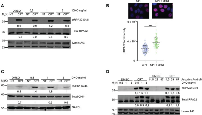
Methods: Here, we used a hydrophilic extract of a new tomato genotype (named DHO) in order to modulate the Camptothecin (CPT) DNA damage response.

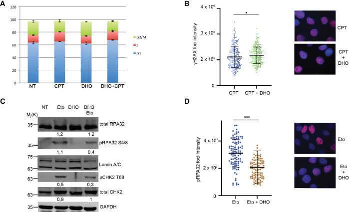
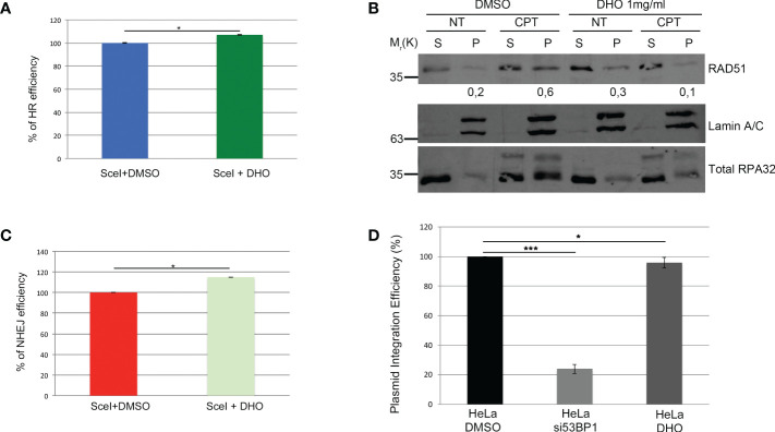
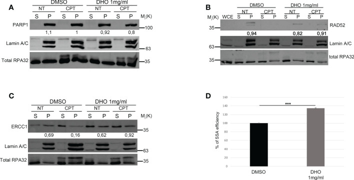
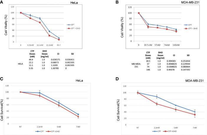
Results: We demonstrated increased phosphorylation of Replication Protein A 32 Serine 4/8 (RPA32 S4/8) protein in HeLa cells treated with the CPT in combination with DHO extract with respect to CPT alone. Moreover, we pointed out a change in HR intermediates resolution from Gene Conversion to Single Strand Annealing through the modified DNA repair protein RAD52 homolog (RAD52), DNA excision repair protein ERCC-1 (ERCC1) chromatin loading in response to DHO extract, and CPT co-treatment, with respect to the vehicle. Finally, we showed an increased sensitivity of HeLa cell lines to DHO extract and CPT co-treatment suggesting a possible mechanism for increasing the efficiency of cancer therapy.

Discussion: We described the potential role of DHO extract in the modulation of DNA repair, in response to Camptothecin treatment (CPT), favoring an increased sensitivity of HeLa cell lines to topoisomerase inhibitor therapy.

Keywords: DNA damage response; DNA repair; Solanum lycopersicum; homologous recombination; single strand annealing.


Conflict of interest statement

The authors declare that the research was conducted in the absence of any commercial or financial relationships that could be construed as a potential conflict of interest.

Figures

Figure 1
Figure 1
DHO hydrophilic extract increases the RPA32 S4/8 phosphorylation in response to CPT respect to CPT alone. (A) Western blot analysis of HeLa cells pre-treated, for one hour, with different concentrations (0.5, 1, 3 mg/ml) of DHO extract and DMSO followed by a treatment with 1 μM CPT for additional two hours. Total RPA32 and Lamin A/C are loading as control of pRPA32 S4/8 and total RPA32, respective. The values obtained are reported in the blot images. (B) Immunofluorescence analysis of pRPA32 S4/8 foci intensity in HeLa cells pretreated for one hour with DHO 1mg/ml or DMSO, followed incubation by 1 μM CPT for additional two hours. The histogram reports three independent experiments with standard deviations (SD). Statistically significant differences are indicated with: ***significant (P < 0.001). (C) Western blot analysis of HeLa cells treat with DHO with different concentrations (0.5, 1, 3 mg/ml) and CTP (1 µM) alone and in combination. CHK1 S345 phosphorylation (pCHK1 S345). Total CHK1 and GAPDH are loading as control of pCHK1 S345 and total CHK1, respective. The values obtained are reported in the blot images. (D) Western blot analysis of HeLa cells pre-treated for one hour with different DHO (0.5, 1, 3 mg/ml) or Ascorbic Acid (14.5, 29 and 87 μM) concentrations followed by two hours of CPT treatment. pRPA32 S4/8 was normalized as indicated above.
Figure 2
Figure 2
DHO hydrophilic extract do not alter cell cycle distribution anh H2AX activation (A) Cell cycle profile of HeLa cells pre-treated with 1 mg/ml of DHO or vehicle (DMSO) for one hours followed by incubation with CPT (1 µM) for additional two hours, was analysed through flow cytometry upon propidium iodide (PI) staining. (B) Immunofluorescence analysis of γH2AX foci intensity in HeLa cells pretreated for one hour with DHO 1mg/ml or vehicle (DMSO), followed incubation by 1 μM CPT for additional two hours. The histogram reports the mean of three independent experiments with standard deviations (SD). Statistically significant differences are indicated with: *significant (P < 0.05). (C) Western blot analysis of HeLa cells pre-treated for one hour with DHO at 1 mg/ml or vehicle (DMSO), followed by two hours of Etoposide (20 μM) incubation. RPA S4/8 and pCHK1 S345 or vehicle (DMSO), followed by two hours of Etoposide (20 μM) incubation. RPA S4/8 and pCHK1 S345 were normalized to those of each respective total RPA32 and CHK1 controls. (D) Immunofluorescence analysis of pRPA32 S4/8 foci intensity in HeLa cells pre-treated with 1mg/ml of DHO followed by incubation with Etoposide 20 µM for additional two hours. The histogram reports the mean of three independent experiments with standard deviations (SD). Statistically significant differences are indicated with: ***significant (P < 0.001).
Figure 3
Figure 3
DHO hydrophilic extract impairs RAD51 chromatin loading (A) HeLa DR-GFP cells were transfected with the coding plasmid for the SceI restriction enzyme in presence of 1 mg/ml of DHO or vehicle (DMSO) and incubated for 48 hours. FACS analysis measurement of GFP levels was used to calculate %HR frequency compared with DMSO alone which was set as 100% (control). Data represent the mean % ± SD obtained from three independent experiments. Statistically significant differences are indicated with: *significant (P < 0.05). (B) Chromatin enhriched purification of HeLa cells pretreated or not with the DHO extract at 1 mg/ml followed by 1 μM CPT treatment for additional two hours. Cells were then lysed to obtain a soluble (S) and a chromatin-enriched (P, as pellet) fraction. Western blotting of RAD51 was performed to analyse the loading onto chromatin of the indicated proteins, involved in the cell response to DNA damage. Total RPA32 and Lamin A/C were used as controls of the supernatant or the chromatin-enriched fraction, respectively. (C) HeLa pimEJ5-GFP cells were transfected with the plasmid encoding the SceI restriction enzyme followed by incubation with 1 mg/ml of DHO extract or vehicle (DMSO) for 48 hours followed by FACS analysis measurement of GFP levels to calculate %NHEJ frequency compared with control cells which were set as 100%. Data represent the mean ± SD obtained from three independent experiments. Statistically significant differences are indicated with: *significant (P < 0.05). (D) HeLa cells were transfected with the pCMV-His empty vector linearized with the ApaI restriction enzyme and pEGFP-C1 in presence of siCTR or si53BP1 followed by incubation with the vehicle (DMSO) or 1 mg/ml DHO hydrophilic extract for 15 days and stained with 2% (w/v) crystal violet solution. Data represent the mean % ± SD obtained from three independent experiments. Statistically significant differences are indicated with: *significant (P < 0.05), ***significant (P < 0.001).
Figure 4
Figure 4
DHO hydrophilic extract increases the Single Strand Annealing activity in response to CPT treatment (A) Chromatin enhriched purification of HeLa cells pretreated or not with the DHO extract at 1 mg/ml followed by 1 μM CPT treatment for additional two hours. Cells were then lysed to obtain a soluble (S) and a chromatin-enriched (P, as pellet) fraction. Western blotting was performed to analyse the loading onto chromatin of the Poly (ADP-ribose) polymerase (PARP1) protein, involved in the cell response to DNA damage. Total RPA32 and Lamin A/C were used as controls of the supernatant or the chromatin-enriched fraction, respectively. (B) Chromatin enhriched purification of HeLa cells performed as previously for the analysis of RAD52 chromatin loading. (C) Chromatin enhiched purification of HeLa cells was performed as previously described followed by incubation with ERCC1 antibody. (D) HeLa hprtSAGFP cells were transfected with the plasmid encoding the SceI restriction enzyme followed by incubation with 1 mg/ml of DHO extract or vehicle (DMSO) for 48 hours followed by FACS analysis measurement of GFP levels to calculate %SSA frequency compared with control cells which were set as 100%. Data represent the mean % ± SD. obtained from three independent experiments. Statistically significant differences are indicated with: ***significant (P < 0.001).
Figure 5
Figure 5
Synergistic effect of DHO-CPT combination on different cancer cell lines (A) HeLa cells were pretreated with 1 mg/ml of DHO hydrophilic extract or vehicle (DMSO) followed by different doses of CPT for additional 72 hours and cell viability was analyzed by MTS assay. Results represent the means and SD of three independent experiments, each conducted in triplicate, and are expressed as percentages of cell viability, calculated with respect to the control cells treated with DMSO alone. Combination index was calculated as described in matherial and methods. (B) MDA-MB-231 cells were pretreated with 1mg/ml of DHO or vehicle followed by incubation with different doses of CPT for additional 72 hours and cell viability assay was performed as previously. (C) HeLa cells were pretreated with DHO at 1mg/ml for one hour followed by treatment with CPT at indicated concentrations; at the end of ten days of incubations, Hela cells were stained with crystal violet. (D) MDA-MB-231 cells were incubated with DHO, as described previously, for the cell viabilty assay and cultured for ten days followed by staining with the cristal violet.

Similar articles

Cited by

References

    1. Hanahan D, Weinberg RA, Adams JM, Cory S, Aguirre-Ghiso JA, Ahmed Z, et al. . Hallmarks of cancer: the next generation. Cell (2011) 144:646–74. doi: 10.1016/j.cell.2011.02.013 - DOI - PubMed
    1. Lieber MR. The mechanism of human nonhomologous DNA end joining. J Biol Chem (2008) 283:1–5. doi: 10.1074/jbc.R700039200 - DOI - PubMed
    1. San Filippo J, Sung P, Klein H. Mechanism of eukaryotic homologous recombination. Annu Rev Biochem (2008) 77:229–57. doi: 10.1146/annurev.biochem.77.061306.125255 - DOI - PubMed
    1. Ceccaldi R, Rondinelli B, D’Andrea A. Repair pathway choices and consequences at the double-strand break. Trends Cell Biol (2016) 26:52–64. doi: 10.1016/J.TCB.2015.07.009 - DOI - PMC - PubMed
    1. Symington LS. Mechanism and regulation of DNA end resection in eukaryotes. Crit Rev Biochem Mol Biol (2016) 51:195–212. doi: 10.3109/10409238.2016.1172552 - DOI - PMC - PubMed