Review
. 2023 Jun 20:10:1212314.
doi: 10.3389/fmed.2023.1212314. eCollection 2023.

Management of keratoconus: an updated review

Affiliations
Review

Management of keratoconus: an updated review

Rashmi Deshmukh et al. Front Med (Lausanne). .

Abstract

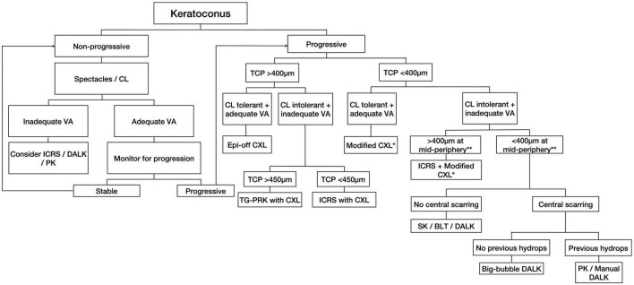
Keratoconus is the most common corneal ectatic disorder. It is characterized by progressive corneal thinning with resultant irregular astigmatism and myopia. Its prevalence has been estimated at 1:375 to 1:2,000 people globally, with a considerably higher rate in the younger populations. Over the past two decades, there was a paradigm shift in the management of keratoconus. The treatment has expanded significantly from conservative management (e.g., spectacles and contact lenses wear) and penetrating keratoplasty to many other therapeutic and refractive modalities, including corneal cross-linking (with various protocols/techniques), combined CXL-keratorefractive surgeries, intracorneal ring segments, anterior lamellar keratoplasty, and more recently, Bowman's layer transplantation, stromal keratophakia, and stromal regeneration. Several recent large genome-wide association studies (GWAS) have identified important genetic mutations relevant to keratoconus, facilitating the development of potential gene therapy targeting keratoconus and halting the disease progression. In addition, attempts have been made to leverage the power of artificial intelligence-assisted algorithms in enabling earlier detection and progression prediction in keratoconus. In this review, we provide a comprehensive overview of the current and emerging treatment of keratoconus and propose a treatment algorithm for systematically guiding the management of this common clinical entity.

Keywords: artificial intelligence; contact lens; cornea; corneal cross-linking; corneal transplant; intracorneal ring segment; keratoconus; refractive surgery.


Conflict of interest statement

The authors declare that the research was conducted in the absence of any commercial or financial relationships that could be construed as a potential conflict of interest.

Figures

FIGURE 1
FIGURE 1
A proposed treatment algorithm for guiding the management of keratoconus. CL, contact lens; VA, visual acuity; ICRS, intracorneal ring segments; DALK, deep anterior lamellar keratoplasty; PK, penetrating keratoplasty; TCP, thinnest corneal pachymetry; CXL, corneal cross-linking; TG-PRK, topographic guided-photorefractive keratectomy; SK, stromal keratophakia; BLT, Bowman’s layer transplantation; DM, Descemet membrane. *Modified CXL includes transepithelial, iontophoresis-assisted, lenticule-assisted, CL-assisted, and adapted fluence CXL. **Corneal thickness at the mid-periphery/tunneling site for ICRS implantation.
FIGURE 2
FIGURE 2
Infectious keratitis following epithelium-off corneal cross-linking for progressive keratoconus. (A) Active infection. (B) Resolved infection with a visual debilitating corneal scar.
FIGURE 3
FIGURE 3
A case of intracorneal ring segments (ICRS) implantation for treating stable moderate keratoconus. The (A) preoperative and (B) postoperative corneal topography demonstrates an improvement in the regularity of the cornea following ICRS.
FIGURE 4
FIGURE 4
Surgical planning of Keraring intracorneal ring segments implantation based on preoperative refraction and tomography.
FIGURE 5
FIGURE 5
Slit-lamp photograph demonstrating an extrusion of intracorneal ring segments (black arrows), which is a well-recognized complication.
FIGURE 6
FIGURE 6
A case of corneal cross-linking (CXL) plus [with wavefront transepithelial photorefractive keratectomy (tPRK) followed by simultaneous same-day conventional CXL] for treating moderate progressive keratoconus. (A) Preoperative corneal topography demonstrating a significant irregular astigmatism [with high inferior-superior (IS) asymmetry] and a corrected-distance-visual-acuity (CDVA) of 0.5 logMAR. (B) Postoperative corneal topography demonstrating a significant improvement in irregular astigmatism and CDVA to 0.0 logMAR following the treatment. (C) The ablation map demonstrating the setting of the tPRK used in this patient.
FIGURE 7
FIGURE 7
Anterior segment optical coherence tomography (AS-OCT) images demonstrating the outcome of implanted autologous adipose derived adult stem cells (ADASCs) in advanced keratoconus. (A) An ASOCT image showing the effect of the cellular therapy of the corneal stroma by an intrastromal implantation of autologous ADASCs in a patient with advanced keratoconus at 6 months post-treatment. Observe the hyperreflective band of neo-collagen (around 15 μm thickness) at the level of the stromal pocket (arrows). (B) An ASOCT image showing the corneal stromal enhancement by an intrastromal implantation of a decellularized lamina of human corneal stroma (arrows) colonized with autologous ADASC in a patient with advanced keratoconus at 12 months post-treatment.

References

    1. Wyman A. Benedict Duddell: pioneer oculist of the 18th century. J R Soc Med. (1992) 85:412–5. - PMC - PubMed
    1. Grzybowski A, McGhee C. The early history of keratoconus prior to Nottingham’s landmark 1854 treatise on conical cornea: a review. Clin Exp Optom. (2013) 96:140–5. 10.1111/cxo.12035 - DOI - PubMed
    1. Gokul A, Patel D, McGhee CN. John Nottingham’s 1854 landmark treatise on conical cornea considered in the context of the current knowledge of keratoconus. Cornea. (2016) 35:673–8. 10.1097/ICO.0000000000000801 - DOI - PubMed
    1. Ferrari G, Rama P. The keratoconus enigma: a review with emphasis on pathogenesis. Ocul Surf. (2020) 18:363–73. 10.1016/j.jtos.2020.03.006 - DOI - PubMed
    1. Gorskova E, Sevost’ianov E. Epidemiology of keratoconus in the Urals. Vestn Oftalmol. (1998) 114:38–40. - PubMed