Review
. 2023 Sep 1;24(9):589-601.
doi: 10.2459/JCM.0000000000001484. Epub 2023 May 29.

Diagnosis and treatment of fetal and pediatric age patients (0-12 years) with Wolff-Parkinson-White syndrome and atrioventricular accessory pathways

Affiliations
Review

Diagnosis and treatment of fetal and pediatric age patients (0-12 years) with Wolff-Parkinson-White syndrome and atrioventricular accessory pathways

Loira Leoni et al. J Cardiovasc Med (Hagerstown). .

Abstract

Overt or concealed accessory pathways are the anatomic substrates of ventricular preexcitation (VP), Wolff-Parkinson-White syndrome (WPW) and paroxysmal supraventricular tachycardia (PSVT). These arrhythmias are commonly observed in pediatric age. PSVT may occur at any age, from fetus to adulthood, and its symptoms range from none to syncope or heart failure. VP too can range from no symptoms to sudden cardiac death. Therefore, these arrhythmias frequently need risk stratification, electrophysiologic study, drug or ablation treatment. In this review of the literature, recommendations are given for diagnosis and treatment of fetal and pediatric age (≤12 years) WPW, VP, PSVT, and criteria for sport participation.


Conflict of interest statement

There are no conflicts of interest.

Figures

Fig. 1
Fig. 1
Maternal therapy for in-utero management of fetal tachycardias. SVT, supraventricular tachycardia.
Fig. 2
Fig. 2
Therapeutic strategy in infants with PSVT. See text for further details. PSVT, paroxysmal supraventricular tachycardia; WPW, Wolff–Parkinson–White syndrome.
Fig. 3
Fig. 3
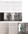
(a) ECG showing VP from a left posterolateral AP. (b) 3D-electroanatomic map of right and left atria (Carto Univu Biosense Webster Inc., diamond bar, CA). From left to right: ablation site local electrogram; left postero-lateral ablation site (red tags), on antero-posterior (middle panel) and left anterior oblique views (left panel). Orange tags: His bundle recording site. Trans-septal approach. (c) ECG showing PSVT (left panel) and 3D-electroanatomic map of right and left atria (Carto Univu Biosense Webster Inc., Diamond Bar, CA) (right panel). Left lateral concealed AP ablation site (red tags, left anterior oblique view). Retrograde transaortic approach. Orange tags: his bundle recording site; light-blue tags: tricuspid annulus. AP, accessory pathway; PSVT, paroxysmal supraventricular tachycardia.
Fig. 4
Fig. 4
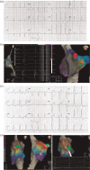
(a) ECG showing VP from a right mid-septal AP. (b) 3D-electroanatomic map of right atrium (EnSite Precision 5.0.1 Abbott Medical). Mid-septal cryoablation site (green tag). Red tags: His bundle recording site. From left to right, left lateral view, ECG and local electrogram at the ablation site showing the disappearance of the delta wave during cryomapping, left anterior oblique view. (c) ECG showing VP from a right antero-septal AP. (d) 3D-electroanatomic map of right atrium (EnSite Precision 5.0.1 Abbott Medical). From left to right, antero-septal cryoablation site (green tags), left and right anterior oblique views. Red tags: his bundle recording site. Last panel shows the disappearance of the delta wave during cryomapping. AP, accessory pathway; PSVT, paroxysmal supraventricular tachycardia; VP, ventricular preexcitation.
Fig. 5
Fig. 5
Flowchart of the management of children with preexcitation syndromes/PSVT after first year of life. See text and Table 2 for further details. A more detailed management algorithm of asymptomatic preexcitation patients can be found in reference 1. Careful assessment of the risk–benefit of ablation based on the patient's weight and size and the site of the accessory pathway. For high-risk asymptomatic patients, the ablation is always a Class II indication. EPS, electrophysiological study; IC, intracardiac; PSVT, paroxysmal supraventricular tachycardia; TE, transesophageal.

References

    1. Cohen MI, Triedman JK, Cannon BC, Davis AM, Drago F, Janousek J, et al. . PACES/HRS expert consensus statement on the management of the asymptomatic young patient with a Wolff–Parkinson–White (WPW, ventricular preexcitation) electrocardiographic pattern. Heart Rhythm 2012; 9:1006–1024. - PubMed
    1. Gillette PC, Freed D, McNamara DG. A proposed autosomal dominant method of inheritance of the Wolff–Parkinson–White syndrome and supraventricular tachycardia. J Pediatr 1978; 93:257–258. - PubMed
    1. Chia BL, Yew FC, Chay SO, Tan AT. Familial Wolff–Parkinson–White syndrome. J Electrocardiol 1982; 15:195–198. - PubMed
    1. MacRae CA, Ghaisas N, Kass S, Donnelly S, Basson CT, Watkins HC, et al. . Familial hypertrophic cardiomyopathy with Wolff–Parkinson–White syndrome maps to a locus on chromosome 7q3. J Clin Invest 1995; 96:1216–1220. - PMC - PubMed
    1. Gollob MH, Green MS, Tang AS, Gollob T, Karibe A, Ali Hassan AS, et al. . Identification of a gene responsible for familial Wolff–Parkinson–White syndrome. N Engl J Med 2001; 344:1823–1831. - PubMed