. 2023 Jul 19;15(28):33362-33372.
doi: 10.1021/acsami.3c04826. Epub 2023 Jul 6.

All-Covalent Nuclease-Resistant and Hydrogel-Tethered DNA Hairpin Probes Map pN Cell Traction Forces

Affiliations

All-Covalent Nuclease-Resistant and Hydrogel-Tethered DNA Hairpin Probes Map pN Cell Traction Forces

Sk Aysha Rashid et al. ACS Appl Mater Interfaces. .

Abstract

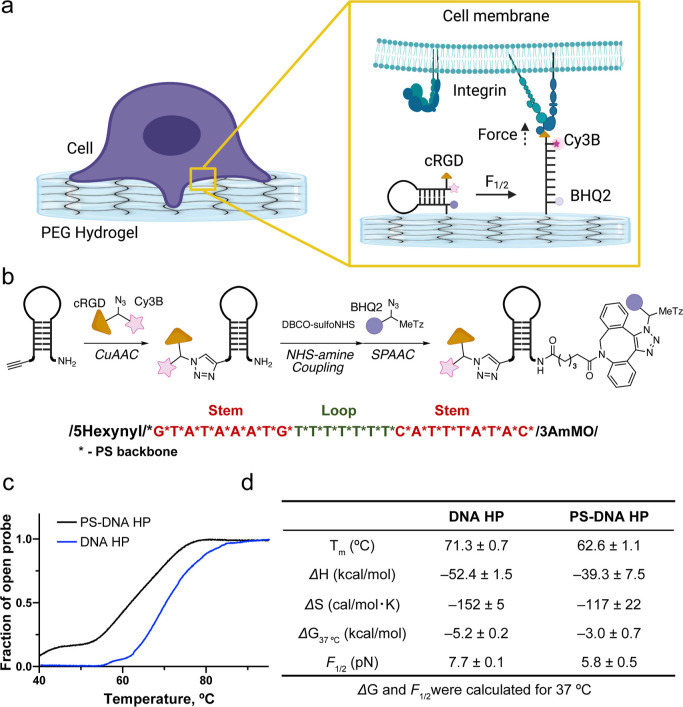
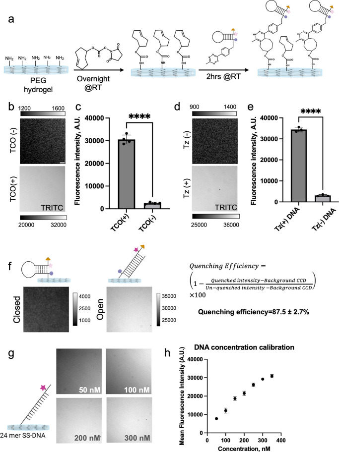
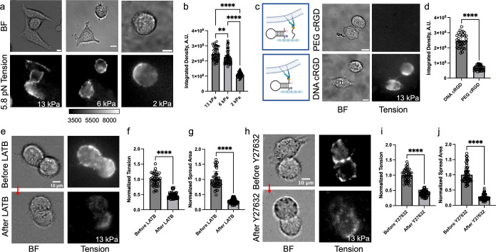
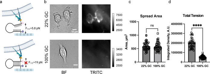
Cells sense and respond to the physical properties of their environment through receptor-mediated signaling, a process known as mechanotransduction, which can modulate critical cellular functions such as proliferation, differentiation, and survival. At the molecular level, cell adhesion receptors, such as integrins, transmit piconewton (pN)-scale forces to the extracellular matrix, and the magnitude of the force plays a critical role in cell signaling. The most sensitive approach to measuring integrin forces involves DNA hairpin-based sensors, which are used to quantify and map forces in living cells. Despite the broad use of DNA hairpin sensors to study a variety of mechanotransduction processes, these sensors are typically anchored to rigid glass slides, which are orders of magnitude stiffer than the extracellular matrix and hence modulate native biological responses. Here, we have developed nuclease-resistant DNA hairpin probes that are all covalently tethered to PEG hydrogels to image cell traction forces on physiologically relevant substrate stiffness. Using HeLa cells as a model cell line, we show that the molecular forces transmitted by integrins are highly sensitive to the bulk modulus of the substrate, and cells cultured on the 6 and 13 kPa gels produced a greater number of hairpin unfolding events compared to the 2 kPa substrates. Tension signals are spatially colocalized with pY118-paxillin, confirming focal adhesion-mediated probe opening. Additionally, we found that integrin forces are greater than 5.8 pN but less than 19 pN on 13 kPa gels. This work provides a general strategy to integrate molecular tension probes into hydrogels, which can better mimic in vivo mechanotransduction.

Keywords: DNA hairpin sensors; DNA tension probes; hydrogel; integrin mechanosensing; mechanobiology; mechanotransduction.


Conflict of interest statement

The authors declare no competing financial interest.

Figures

Figure 1
Figure 1
Preparation and characterization of PEG hydrogels. (a) Schematic design of working principle of hydrogel-tethered DNA hairpin probe. (b) Molecular structure of the hydrogel precursor molecules (tetra PEG-NH2 and tetra PEG-NHS). The PEG polymers used in our work included n = 28, 57, and 114 ethylene glycol monomer units, corresponding to the 5, 10, and 20 kDa polymers, respectively. (c–e) Time-dependent rheology plots of PEG hydrogels synthesized from 5 kDa PEG (c), 10 kDa PEG (d), and 20 kDa PEG (e) precursors. The maximum elastic moduli (Gmax) were estimated when G′ reached a plateau (50 min for 5 and 10 kDa, and 90 min for the 20 kDa precursor). Note that the Gmax values were obtained from triplicate measurements for the 5 and 10 kDa polymers, whereas the Gmax for the 20 kDa polymer was a single measurement, and this was due to the slow polymerization kinetics.
Figure 2
Figure 2
Design, synthesis, and characterization of PS-modified HP probes. (a) Schematic of a hydrogel-tethered DNA hairpin probe and its response to integrin forces. (b) Synthetic scheme for PS-modified DNA HP probes. The red color indicates the self-complementary stem, while the green color indicates the polyT loop. All nucleobases were linked by the PS backbone. (c) Fluorescence thermal melting curves of DNA HP (22% GC, blue) and PS-modified DNA HP (22% GC, black) in 1× PBS. (d) Table of the calculated Tm, ΔG, and F1/2 for DNA HP and PS DNA HP (22% GC) at 37 °C in PBS buffer. The error values represent the standard deviation from three independent melts.
Figure 3
Figure 3
DNA hairpin surface immobilization using TCO-Tz coupling. (a) Schematic showing TCO modification of hydrogel followed by DNA HP conjugation. (b,c) Representative fluorescence images and quantification of TCO-modified hydrogels and control hydrogels following PS-DNA conjugation. (d,e) Representative fluorescence images and quantification of TCO-modified hydrogels following PS-DNA-Tz conjugation and control PS-DNA conjugation. (f) Representative fluorescence images used to determine HP quenching efficiency. As shown schematically, the fully open HP was obtained by hybridization with complement prior to surface tethering. (g,h) DNA concentration-dependent fluorescence images along with quantification show dose-dependent increase in surface density. ****, ***, **, * and ns indicate p < 0.0001, p < 0.001, p < 0.01, p < 0.05, and not significant, respectively, as determined from one-way ANOVA. Error bars show the standard deviation for N > 3, three different sets of surface preparations. Each intensity value was averaged from at least 10 different regions of interest. Scale bar = 10 μm.
Figure 4
Figure 4
DNA HP probes map integrin tension and demonstrate enhanced traction forces in response to the Young’s modulus of the hydrogel. (a) Brightfield (BF) and fluorescence tension maps of HeLa cells cultured on 13, 6, and 2 kPa substrates coated with PS DNA HP probe (22% GC content) for ∼5–6 h. (b) Plot quantifying the total tension signal of individual HeLa cells on the different substrates. (c) Schematic illustration of probe surface and control hydrogel (left) along with BF and fluorescence tension images of HeLa cells cultured on control hydrogel coated with PEG-cRGD and cRGD-lacking PS DNA HP (top) and hydrogel coated with PS DNA HP probe (bottom). (d) Plot quantifying the total tension signal from single cells from the experiments shown in (c). (e–g) BF and tension images of HeLa cells before and after treatment with LatB (20 μM) along with bar graph quantifying single cell spread area and tension signal. Note that the plots were normalized to the before LatB treatment group. (h–j) BF and tension images of HeLa cells before and after treatment with Y27632 (25 μM) drug and bar graph quantifying single cell spread area and tension signal. Note that the plot was normalized to the non-treated cell group. ****, ***, **, *, and ns indicate p < 0.0001, p < 0.001, p < 0.01, p < 0.05, and not significant respectively, as determined from one-way ANOVA. Error bars show the standard deviation for N > 3, where each experiment was averaged from three or more different cell passages with three different sets of surface preparations. For each replicate N, we measured the signal from at least 15 cells. Scale bar = 10 μm.
Figure 5
Figure 5
Forces generated by integrins in HeLa cells are 5.8 pN < F1/2 < 19 pN. (a) Schematic representation showing force-mediated unfolding of 22% GC PS DNA HP probes but not the 100% GC probes. (b) Brightfield and fluorescence tension (Cy3B) images of Hela cells on 5.8 and 19 pN 13 kPa hydrogel surfaces. Images were taken after ∼5–6 h of incubation. (c) Bar graph showing single cell spread area on 22 and 100% GC hairpin surfaces. (d) Bar graphs plotting the integrated fluorescence tension signal of single HeLa cells cultured on hydrogel-tethered 22 and 100% GC content PS DNA HP probes. ****, ***, **, *, and ns indicate p < 0.0001, p < 0.001, p < 0.01, p < 0.05, and not significant, respectively, as determined from one-way ANOVA. Error bars show the standard deviation for N > 3, where each experiment was averaged from three or more different cell passages with three different sets of surface preparations. For Each replicate N, at least 15 cells were quantified. Scale bar = 10 μm.

Similar articles

Cited by

References

    1. Mohammed D.; Versaevel M.; Bruyère C.; Alaimo L.; Luciano M.; Vercruysse E.; Procès A.; Gabriele S. Innovative Tools for Mechanobiology: Unraveling Outside-in and inside-out Mechanotransduction. Front. Bioeng. Biotechnol. 2019, 7, 162.10.3389/fbioe.2019.00162. - DOI - PMC - PubMed
    1. Martino F.; Perestrelo A. R.; Vinarský V.; Pagliari S.; Forte G. Cellular Mechanotransduction: From Tension to Function. Front. Physiol. 2018, 9, 824.10.3389/fphys.2018.00824. - DOI - PMC - PubMed
    1. Jaalouk D. E.; Lammerding J. Mechanotransduction Gone Awry. Nat. Rev. Mol. Cell Biol. 2009, 10, 63–73. 10.1038/nrm2597. - DOI - PMC - PubMed
    1. Chin L.; Xia Y.; Discher D. E.; Janmey P. A. Mechanotransduction in Cancer. Curr. Opin. Chem. Eng. 2016, 11, 77–84. 10.1016/j.coche.2016.01.011. - DOI - PMC - PubMed
    1. Jansen K. A.; Atherton P.; Ballestrem C. Mechanotransduction at the Cell-Matrix Interface. Semin. Cell Dev. Biol. 2017, 71, 75–83. 10.1016/j.semcdb.2017.07.027. - DOI - PubMed