

Reconfigurable Magnetic Liquid Building Blocks for Constructing Artificial Spinal Column Tissues
- PMID: 37409801
- PMCID: PMC10477840
- DOI: 10.1002/advs.202300694
Reconfigurable Magnetic Liquid Building Blocks for Constructing Artificial Spinal Column Tissues
Abstract
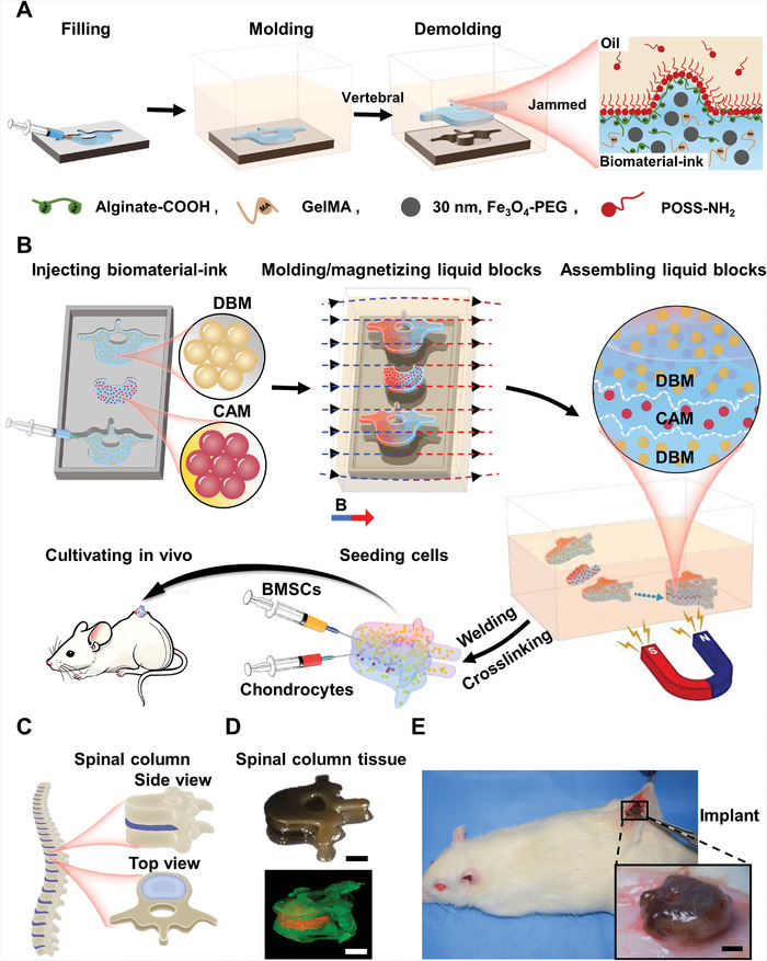
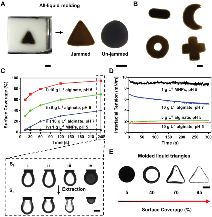
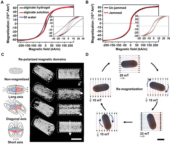
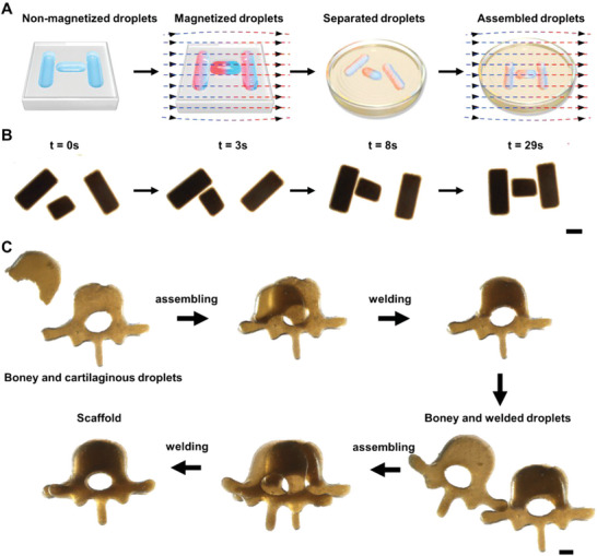
All-liquid molding can be used to transform a liquid into free-form solid constructs, while maintaining internal fluidity. Traditional biological scaffolds, such as cured pre-gels, are normally processed in solid state, sacrificing flowability and permeability. However, it is essential to maintain the fluidity of the scaffold to truly mimic the complexity and heterogeneity of natural human tissues. Here, this work molds an aqueous biomaterial ink into liquid building blocks with rigid shapes while preserving internal fluidity. The molded ink blocks for bone-like vertebrae and cartilaginous-intervertebral-disc shapes, are magnetically manipulated to assemble into hierarchical structures as a scaffold for subsequent spinal column tissue growth. It is also possible to join separate ink blocks by interfacial coalescence, different from bridging solid blocks by interfacial fixation. Generally, aqueous biomaterial inks are molded into shapes with high fidelity by the interfacial jamming of alginate surfactants. The molded liquid blocks can be reconfigured using induced magnetic dipoles, that dictated the magnetic assembly behavior of liquid blocks. The implanted spinal column tissue exhibits a biocompatibility based on in vitro seeding and in vivo cultivating results, showing potential physiological function such as bending of the spinal column.
Keywords: alginate surfactants; all-liquid molding; in vivo cultivated tissues; spinal column structures; structured magnetic droplets.
© 2023 The Authors. Advanced Science published by Wiley-VCH GmbH.
Conflict of interest statement
The authors declare no conflict of interest.
Figures
Similar articles
-
Enhancing cell migration in shape-memory alginate-collagen composite scaffolds: In vitro and ex vivo assessment for intervertebral disc repair.J Biomater Appl. 2015 Apr;29(9):1230-46. doi: 10.1177/0885328214557905. Epub 2014 Nov 6. J Biomater Appl. 2015. PMID: 25376622
-
Self-assembly of aligned tissue-engineered annulus fibrosus and intervertebral disc composite via collagen gel contraction.Tissue Eng Part A. 2010 Apr;16(4):1339-48. doi: 10.1089/ten.TEA.2009.0442. Tissue Eng Part A. 2010. PMID: 19905878 Free PMC article.
-
The significance of biomacromolecule alginate for the 3D printing of hydrogels for biomedical applications.Int J Biol Macromol. 2022 Jul 1;212:561-578. doi: 10.1016/j.ijbiomac.2022.05.157. Epub 2022 May 25. Int J Biol Macromol. 2022. PMID: 35643157 Review.
-
Biocompatible liquid-crystal elastomers mimic the intervertebral disc.J Mech Behav Biomed Mater. 2020 Jul;107:103757. doi: 10.1016/j.jmbbm.2020.103757. Epub 2020 Mar 30. J Mech Behav Biomed Mater. 2020. PMID: 32276188
-
Alginate hydrogels: A potential tissue engineering intervention for intervertebral disc degeneration.J Clin Neurosci. 2023 Jul;113:32-37. doi: 10.1016/j.jocn.2023.05.001. Epub 2023 May 7. J Clin Neurosci. 2023. PMID: 37159956 Review.
Cited by
-
Targeting biofilm infections in humans using small scale robotics.Trends Biotechnol. 2024 Apr;42(4):479-495. doi: 10.1016/j.tibtech.2023.10.004. Epub 2023 Nov 13. Trends Biotechnol. 2024. PMID: 37968157 Free PMC article. Review.
References
-
- Shi S., Russell T. P., Adv. Mater. 2018, 30, 1800714. - PubMed
-
- Forth J., Kim P. Y., Xie G., Liu X., Helms B. A., Russell T. P., Adv. Mater. 2019, 31, 1806370. - PubMed
-
- Ren Y., Liu J., Micromachines 2018, 9, 218. - PubMed
-
- Liu X., Kent N., Ceballos A., Streubel R., Jiang Y., Chai Y., Kim P. Y., Forth J., Hellman F., Shi S., Wang D., Helms B. A., Ashby P. D., Fischer P., Russell T. P., Science 2019, 365, 264. - PubMed
Publication types
MeSH terms
Substances
Grants and funding
- 2019YFA0110500/National Key R&D Program of China
- 2020TQ0329/China Postdoctoral Science Foundation
- 2020YFA0710403/National Key R&D Program of China
- 2021YFA1200403/National Key R&D Program of China
- 82202477/National Natural Science Foundation of China
- 82020108020/National Natural Science Foundation of China
- 82072198/National Natural Science Foundation of China
- 81873941/National Natural Science Foundation of China
- 81701922/National Natural Science Foundation of China
- U.S. Department of Energy, Office of Science
- DE-AC02-05-CH11231/Office of Basic Energy Sciences, Materials Sciences and Engineering Division
- KCTR16/Adaptive Interfacial Assemblies Toward Structuring Liquids program