. 2023 Aug 31;14(4):e0135323.
doi: 10.1128/mbio.01353-23. Epub 2023 Jul 6.

Ergosterol distribution controls surface structure formation and fungal pathogenicity

Affiliations

Ergosterol distribution controls surface structure formation and fungal pathogenicity

Hau Lam Choy et al. mBio. .

Erratum in

Abstract

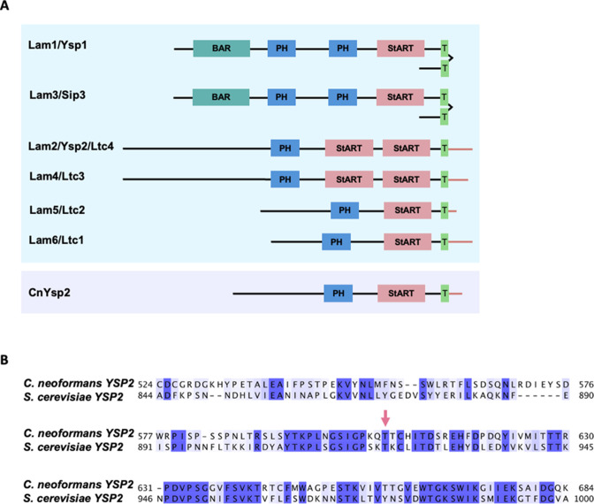
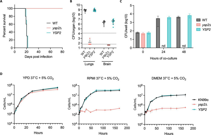
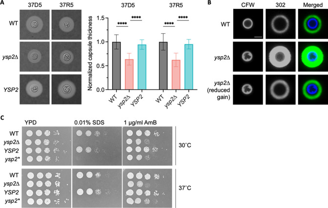
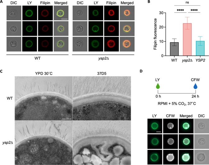
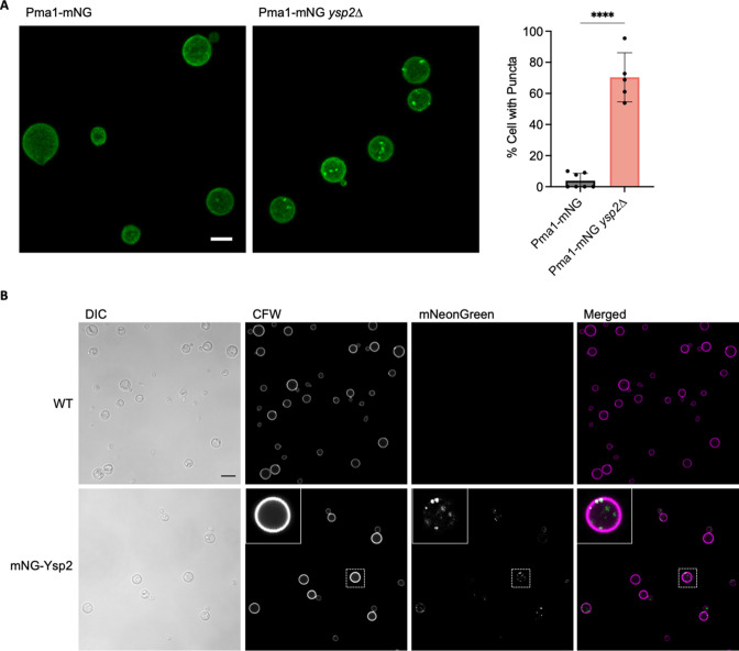
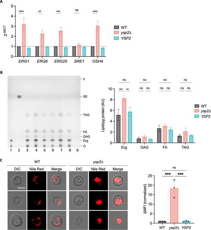
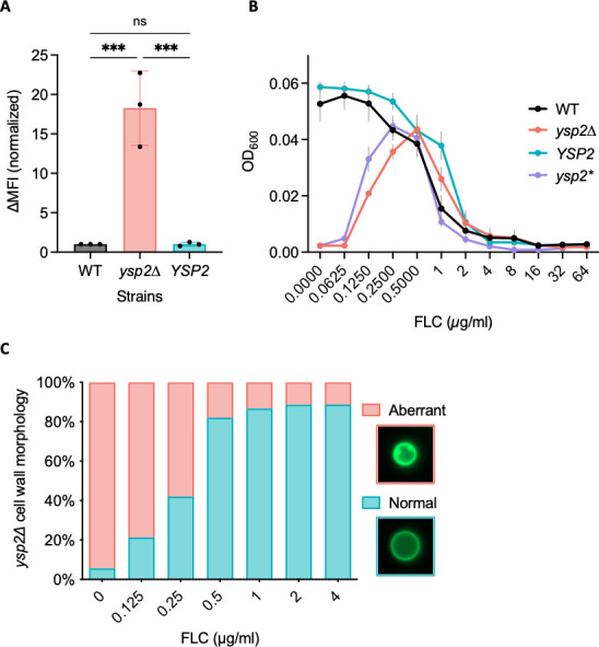
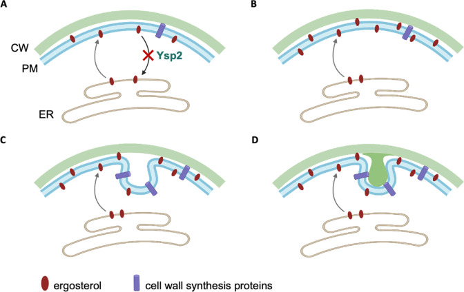
Ergosterol, the major sterol in fungal membranes, is critical for defining membrane fluidity and regulating cellular processes. Although ergosterol synthesis has been well defined in model yeast, little is known about sterol organization in the context of fungal pathogenesis. We identified a retrograde sterol transporter, Ysp2, in the opportunistic fungal pathogen Cryptococcus neoformans. We found that the lack of Ysp2 under host-mimicking conditions leads to abnormal accumulation of ergosterol at the plasma membrane, invagination of the plasma membrane, and malformation of the cell wall, which can be functionally rescued by inhibiting ergosterol synthesis with the antifungal drug fluconazole. We also observed that cells lacking Ysp2 mislocalize the cell surface protein Pma1 and have abnormally thin and permeable capsules. As a result of perturbed ergosterol distribution and its consequences, ysp2∆ cells cannot survive in physiologically relevant environments such as host phagocytes and are dramatically attenuated in virulence. These findings expand our knowledge of cryptococcal biology and underscore the importance of sterol homeostasis in fungal pathogenesis. IMPORTANCE Cryptococcus neoformans is an opportunistic fungal pathogen that kills over 100,000 people worldwide each year. Only three drugs are available to treat cryptococcosis, and these are variously limited by toxicity, availability, cost, and resistance. Ergosterol is the most abundant sterol in fungi and a key component in modulating membrane behavior. Two of the drugs used for cryptococcal infection, amphotericin B and fluconazole, target this lipid and its synthesis, highlighting its importance as a therapeutic target. We discovered a cryptococcal ergosterol transporter, Ysp2, and demonstrated its key roles in multiple aspects of cryptococcal biology and pathogenesis. These studies demonstrate the role of ergosterol homeostasis in C. neoformans virulence, deepen our understanding of a pathway with proven therapeutic importance, and open a new area of study.

Keywords: Cryptococcus neoformans; Ysp2; ergosterol; mycology; sterol transport; virulence.


Conflict of interest statement

The authors declare no conflict of interest.

Figures

Fig 1
Fig 1
Alignment of cryptococcal Ysp2 with S. cerevisiae homologs. (A) LAM family proteins in S. cerevisiae (blue box) and C. neoformans (purple box). Domain abbreviations are Bar, Bin/amphiphysin/RVS; PH, pleckstrin-homology; StART, Steroidogenic Acute Regulatory Transfer-like; and T, transmembrane. (B) Alignment of C. neoformans and S. cerevisiae YSP2 StART-like domains, with CLUSTALX coloring of conserved residues. Pink arrow, residue predicted to make van der Waals contact with ergosterol (22).
Fig 2
Fig 2
Ysp2 is required for virulence and survival in physiological environments. (A) Survival of C57BL/6 mice after intranasal infection with 1.25 × 10⁴ fungal cells, with sacrifice triggered by weight below 80% of initial weight. (B) Lung and brain fungal burdens of mice from Panel A at sacrifice. Top dotted line, initial inoculum; bottom dotted line, limit of detection. (C) In vitro survival of cryptococci. Bone marrow-derived macrophages were co-incubated with the indicated strains (1.5 h, MOI = 0.1) and washed to remove free fungi before lysis at the times shown and assessment of cryptococcal CFU. nd, not detected. Mean ± SD is plotted; results shown are representative of at least two biological replicate experiments. (D) Growth curves in the conditions shown (mean ± SEM of three independent experiments). YPD, yeast extract-peptone-dextrose medium; DMEM, Dulbecco's Modified Eagle Medium.
Fig 3
Fig 3
The ysp2∆ mutant exhibits cell surface defects. (A) Capsule thickness. The indicated strains were grown in host-like conditions [37°C, 5% CO2, with either DMEM (37D5) or RPMI (37R5)], stained with India ink (left), and capsule thickness was measured with ImageJ and normalized to cell radius and WT value (right). Mean ± SD of at least 50 cells per sample are shown. ****P < 0.0001 by one-way analysis of variance. (B) Representative confocal micrographs of the indicated strains after growth in 37R5 for 24  h and staining with CFW (cell wall) and MAb 302 conjugated to Alexa Fluor 488 (capsule). Images in the first and second rows were obtained at the same gain and intensity settings; in the third row, confocal gain of the α-capsule Ab channel was reduced (reduced gain). All images are to the same scale; bar, 5  µm. (C) Serial 10-fold dilutions of the indicated strains in the conditions shown. ysp2*, inactivated mutant (T606D) with abrogated sterol-binding activity.
Fig 4
Fig 4
The ysp2∆ mutant exhibits malformations of the cell wall and plasma membrane. (A) Representative cells that were grown in 37R5 for 24 h and stained with Lucifer Yellow (LY) for cell wall and filipin for non-esterified sterols. All images are to the same scale; bar, 5 µm. DIC, differential interference contrast. (B) Filipin fluorescence (mean ± SD of mean gray value for at least 50 cells per sample). ****P < 0.0001 by one-way analysis of variance. (C) Transmission electron micrographs of cells grown in 37D5. All images are to the same scale; bar, 500 nm. (D) Representative ysp2∆ cells that were stained with LY, grown in 37R5 for 24 h, and stained with CFW. All images are to the same scale; bar, 5 µm.
Fig 5
Fig 5
Protein localization imaged by confocal microscopy. (A) Pma1-mNeonGreen (Pma1-mNG) expressed in WT and ysp2∆ cells. Images, maximum intensity projections that sum Z-stacks. Both are to the same scale; bar, 5 µm. Plot, percent of cells with fluorescent puncta (mean ± SD based on at least 50 cells across five to seven image fields). ****P < 0.0001 by Student’s t test. (B) Fluorescence and DIC images of WT cells alone (top) or the same cells expressing mNeonGreen-Ysp2 (mNG-Ysp2, bottom). Inset, an example cell (boxed) enlarged threefold. Bar, 5 µm.
Fig 6
Fig 6
Sterol content, synthesis, and distribution. (A) Expression of ergosterol-related genes, measured by RT-qPCR and normalized to ACT1 expression and WT values. The mean ± SEM of three independent experiments is shown. ns, not significant. ****P < 0.0001, ***P < 0.001, and **P < 0.01 by one-way analysis of variance. (B) Lipid profile assessed by thin layer chromatography. Left, a representative thin layer chromatograph, showing two independent biological replicate sample sets. Lane 1, 5 µg/mL ergosterol standard; lane 2, 10 µg/mL cholesteryl oleate standard; lanes 3 and 6, WT; lanes 4 and 7, ysp2∆; lanes 5 and 8, YSP2; 9, vehicle control. O, origin; F, solvent front; Erg, ergosterol; DAG, diacylglycerols; FA, fatty acids; TAG, triacylglycerols; SE, steryl esters. Right, relative lipid abundance. Mean ± SEM of cellular lipids [identified as in reference (41)], measured by densitometry and normalized to total protein (Fig. S6A) are shown for four independent experiments. ***P < 0.001, and **P < 0.01 by one-way analysis of variance. (C) Nile Red staining. Left, representative fluorescence images of WT and mutant strains. All images are to the same scale; bar, 5 µm. Right, ∆MFI (change in median fluorescent intensity) from flow cytometry profiles of the indicated strains. Mean ± SD, normalized to WT, is shown for three independent experiments. **P < 0.01 by one-way analysis of variance.
Fig 7
Fig 7
Antifungal drug interactions with ysp2∆ cells. (A) Amphotericin B-Cy5 binding measured by flow cytometry. Mean ± SD of change in median fluorescent intensity (∆MFI), normalized to WT, is shown for three independent experiments. ns, not significant; ***P < 0.001 by one-way analysis of variance. (B) Mutant response to fluconazole (FLC). Cells were grown in 37R5 with FLC as indicated and OD600 measured at 48 h. Mean ± SEM of three independent experiments is shown. ysp2*, inactivated mutant. (C) Cells were grown in 37R5, and the fraction with aberrant cell walls was quantified at 24 h. Seventy cells were scored per condition.
Fig 8
Fig 8
Model of Ysp2 function and how its absence influences plasma membrane and cell wall morphology. CW, cell wall.

Update of

Similar articles

Cited by

References

    1. Rajasingham R, Govender NP, Jordan A, Loyse A, Shroufi A, Denning DW, Meya DB, Chiller TM, Boulware DR. 2022. The global burden of HIV-associated cryptococcal infection in adults in 2020: a modelling analysis. Lancet Infect Dis 22:1748–1755. doi:10.1016/S1473-3099(22)00499-6 - DOI - PMC - PubMed
    1. Ballou ER, Johnston SA. 2017. The cause and effect of Cryptococcus interactions with the host. Curr Opin Microbiol 40:88–94. doi:10.1016/j.mib.2017.10.012 - DOI - PubMed
    1. Sabiiti W, May RC. 2012. Mechanisms of infection by the human fungal pathogen Cryptococcus neoformans. Future Microbiol 7:1297–1313. doi:10.2217/fmb.12.102 - DOI - PubMed
    1. O’Meara TR, Alspaugh JA. 2012. The Cryptococcus neoformans capsule: a sword and a shield. Clin Microbiol Rev 25:387–408. doi:10.1128/CMR.00001-12 - DOI - PMC - PubMed
    1. Rodrigues ML. 2018. The multifunctional fungal ergosterol. mBio 9:e01755-18. doi:10.1128/mBio.01755-18 - DOI - PMC - PubMed

Publication types