. 2023 Aug 31;14(4):e0083323.
doi: 10.1128/mbio.00833-23. Epub 2023 Jul 6.

Direct interaction of small non-coding RNAs CjNC140 and CjNC110 optimizes expression of key pathogenic phenotypes of Campylobacter jejuni

Affiliations

Direct interaction of small non-coding RNAs CjNC140 and CjNC110 optimizes expression of key pathogenic phenotypes of Campylobacter jejuni

Brandon Ruddell et al. mBio. .

Abstract

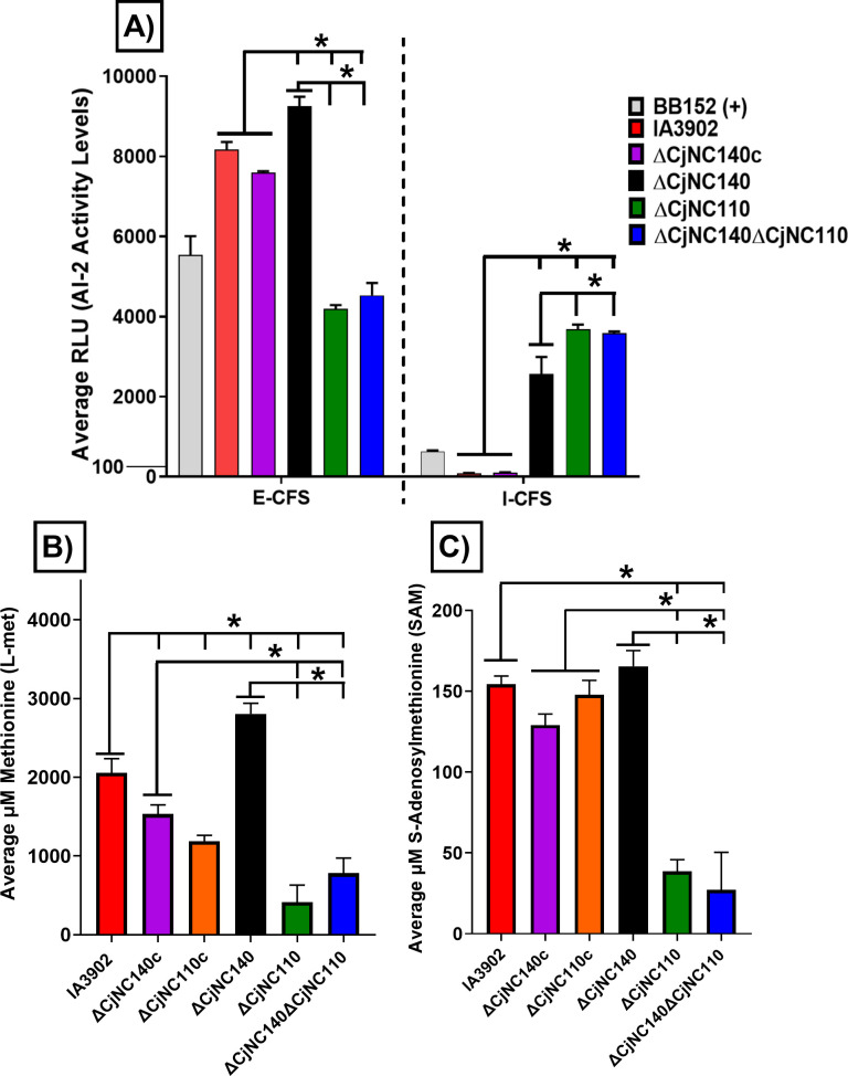
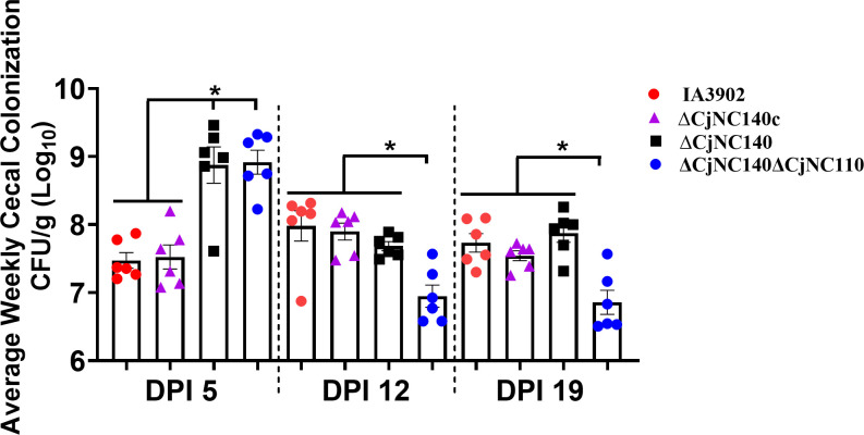
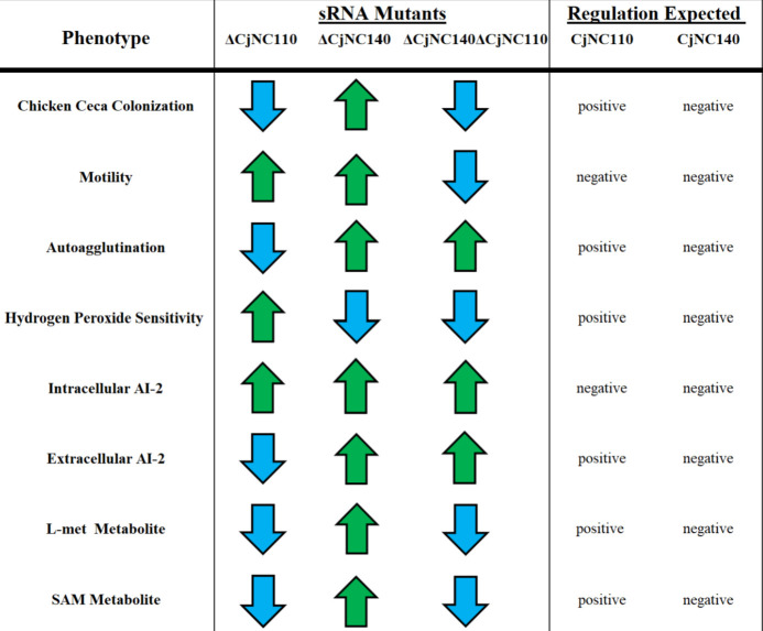
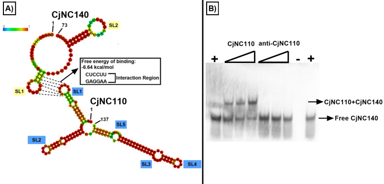
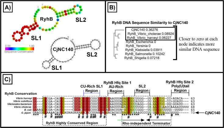
Small non-coding RNAs (sRNAs) are important players in modulating gene expression in bacterial pathogens, but their functions are largely undetermined in Campylobacter jejuni, an important cause of foodborne gastroenteritis in humans. In this study, we elucidated the functions of sRNA CjNC140 and its interaction with CjNC110, a previously characterized sRNA involved in the regulation of several virulence phenotypes of C. jejuni. Inactivation of CjNC140 increased motility, autoagglutination, L-methionine concentration, autoinducer-2 production, hydrogen peroxide resistance, and early chicken colonization, indicating a primarily inhibitory role of CjNC140 for these phenotypes. Apart from motility, all these effects directly contrasted the previously demonstrated positive regulation by CjNC110, suggesting that CjNC110 and CjNC140 operate in an opposite manner to modulate physiologic processes in C. jejuni. RNAseq and northern blotting further demonstrated that expression of CjNC140 increased in the absence of CjNC110, while expression of CjNC110 decreased in the absence of CjNC140, suggesting a possibility of their direct interaction. Indeed, electrophoretic mobility shift assay demonstrated a direct binding between the two sRNAs via GA- (CjNC110) and CU- (CjNC140) rich stem-loops. Additionally, RNAseq and follow-up experiments identified that CjNC140 positively regulates p19, which encodes a key iron uptake transporter in Campylobacter. Furthermore, computational analysis revealed both CjNC140 and CjNC110 are highly conserved in C. jejuni, and the predicted secondary structures support CjNC140 as a functional homolog of the iron regulatory sRNA, RyhB. These findings establish CjNC140 and CjNC110 as a key checks-and- balances mechanism in maintaining homeostasis of gene expression and optimizing phenotypes critical for C. jejuni pathobiology. IMPORTANCE Gene regulation is critical to all aspects of pathogenesis of bacterial disease, and small non-coding RNAs (sRNAs) represent a new frontier in gene regulation of bacteria. In Campylobacter jejuni, the role of sRNAs remains largely unexplored. Here, we investigate the role of two highly conserved sRNAs, CjNC110 and CjNC140, and demonstrate that CjNC140 displays a primarily inhibitory role in contrast to a primarily activating role for CjNC110 for several key virulence-associated phenotypes. Our results also revealed that the sRNA regulatory pathway is intertwined with the iron uptake system, another virulence mechanism critical for in vivo colonization. These findings open a new direction for understanding C. jejuni pathobiology and identify potential targets for intervention for this major foodborne pathogen.

Keywords: Campylobacter; iron homeostasis; post-transcriptional regulation; small non-coding RNAs; sponge RNAs.


Conflict of interest statement

The authors declare no conflict of interest.

Figures

Fig 1
Fig 1
ΔCjNC110 increases the expression of CjNC140, and ΔCjNC140 decreases the expression of CjNC110 relative to IA3902 wild type. (A and B) Northern blot detection of CjNC140. (C and D) Northern blot detection of CjNC110. All strains used and corresponding lanes are indicated in the legend at the top of the image; L, prestained RNA ladder (200b). Cultures for RNA extraction were collected at exponential phase of growth (3 hours) and stationary phase of growth (12 hours) from three separate replicates per strain tested. Northern blots were performed using 12 µg of total RNA. The arrows indicate the dominant band(s) detected at each time point (black boxes), which correspond to CjNC140 (73b; top) and CjNC110 (137b; bottom), as demonstrated previously in Campylobacter jejuni (12, 20). Statistical analysis of band intensity was conducted using one-way analysis of variance. Significance is denoted by "*" when comparing CjNC140 expression between wild-type and ΔCjNC110 or CjNC110 expression between wild type and ΔCjNC140 (black lines).
Fig 2
Fig 2
∆CjNC140 increases both (A) motility and (B) autoagglutination ability and decreases (C) hydrogen peroxide (H2O2) sensitivity when compared to IA3902 wild type (mean ± SEM at 24 hours). Colored bars indicate the average of each strain tested using at least three technical replicates from three independent studies. (A) Motility assays were performed using 0.4% semi-solid agar and measured (mm) by taking the outermost zone of swarming activity. (B) Autoagglutination ability was measured by calculating the optical density (A600) of the supernatant; a lower A600 value indicates increased autoagglutination ability. (C) The zone of sensitivity to H2O2 was measured (mm) by taking the outermost zone of growth inhibition. For statistical analysis, one-way or two-way analysis of variance was performed for each assay when appropriate. Significance (P < 0.05) is denoted by "*" when comparing respective strains (black lines).
Fig 3
Fig 3
ΔCjNC140 increases extracellular (E-CFS) and intracellular cell free supernatant (I-CFS) autoinducer-2 (AI-2) levels, and L-methionine (L-met) when compared to IA3902 wild type (mean ± SEM). Colored bars indicate the average of each strain tested using three technical replicates from three independent experiments. (A) Relative light units (RLU) corresponding to AI-2 activity levels at 12 hours. The average RLU of Vibrio harveyi strain BB152 was used as an internal positive control for relative comparison. For statistical analysis, two-way analysis of variance (ANOVA) using repeated measures with Sidak’s multiple comparison test was performed for each AI-2 assay; no comparison is made for the positive control. (B and C) TR-FRET fluorescence assays measuring L-met and S-adenosylmethionine (SAM) metabolite concentrations. Cultures were collected for cell pellets and normalized to 25 mg to extract intracellular metabolites. Standard curves were generated to calculate metabolite concentration (L-met or SAM). For statistical analysis, one-way ANOVA with Tukey’s multiple comparison test was performed for each metabolite assay. Significance (P < 0.05) is denoted by "*" when comparing respective strains (black lines).
Fig 4
Fig 4
ΔCjNC140 increases chick cecal colonization levels at DPI five when compared to IA3902 wild type (mean ± SEM). Each dot represents an individual bird, and each color indicates the strain utilized, as indicated on the right; results collected from a single study. Each bar represents the average colony forming units (CFU) per g (Log10), with a minimum of six birds per strain each week. Significant differences in colonization between strains were tested using one-way analysis of variance (ANOVA) with Tukey’s multiple comparison test at each DPI independently. Significance (P < 0.05) is denoted by "*" when comparing respective strains (black lines). A Shapiro-Wilk test was performed for each day post-inoculation (DPI) to confirm normal distribution and adequate sample size (N) representation per group (P values >0.05), and a Brown–Forsythe test was performed at each DPI to confirm the equal variances assumption (P values >0.05). Inoculum suspension was on average 5×10⁷ CFU/mL for each respective strain, and one-way ANOVA did not detect a statistical difference in inoculum between strains (P > 0.05).
Fig 5
Fig 5
Summary of virulence and colonization determinants as regulated by CjNC140 and CjNC110. Phenotypic trends for each small RNA mutant background relative to IA3902 wild type are indicated by arrows (increase, green arrow; decrease, blue arrow). The post-transcriptional regulation expected (positive, activation; negative, repression) by CjNC110 and CjNC140 in IA3902 wild type for each phenotype is indicated to the right. Results for chicken colonization in CjNC110 are summarized from Kreuder et al. (20).
Fig 6
Fig 6
Computational and molecular evidence demonstrates that CjNC140 and CjNC110 directly interact with each other. (A) Computational analysis of the RNA sequence of both CjNC140 (73b) and CjNC110 (137b) were input within RNAfold (28) to reveal the secondary structure of both sRNAs using minimum free energy and partition functions. The base-pair probability of formation for each individual sRNA is colored (top left, red=high; blue=low); predicted stem-loop (SL) structures are labeled in blue for CjNC110 and yellow for CjNC140. RNAup (28) was used to predict interactions along with the total free energy of binding (kcal/mol) between the sRNAs. The interaction was predicted to occur at SL1 of each sRNA corresponding to RNA bases AAGGAG (CjNC110) and CUCCUU (CjNC140). GraphPad was used to graph the interacting bases (dashed lines) using the results from RNAfold and RNAup. (B) Electrophoretic mobility shift assay detecting bound and free biotin-labeled CjNC140 identifies CjNC140:CjNC110 duplex formation and confirms that both sRNAs interact at their respective SL1s in vitro. The positive (+) and negative controls (−) are indicated, as well as the gradient shift of increasing CjNC110 RNA oligos (0–200 nM). Arrows indicate either bound (top) or free biotin-labeled CjNC140 (bottom).
Fig 7
Fig 7
Translational fusion and electrophoretic mobility shift assays (EMSA) demonstrate regulatory targeting and stabilization of p19 mRNA by CjNC140. (A) Illustration of PMW10 shuttle plasmid series carrying 3902gfpcj-T (positive control) and corresponding fusions, 3902gfpcj-T::p19, 3902gfpcj-T::p19 M1 (point mutation), and 3902gfpcj-T::murD (negative control). Black arrows represent the codon-optimized 3902gfpcj gene sequence (34), and purple arrows represent 3902gfpcj gene fusions. Blue arrows illustrate the porA promoter PporA, and the lollipop shape represents the Rho-independent terminator TporA. All constructs were tested in ∆CjNC140c as the sRNA is highly expressed in this strain. (B) Translational fusion assays reveal regulatory activation of p19 mRNA by CjNC140, as indicated by increased fluorescence signal of PMW10::3902gfpcj-T::p19 (colored bars indicate mean ± SEM at 3 hours). The fluorescence signal is in relative light units (RLU) and corrected for baseline Mueller–Hinton broth background. Fluorescence values were measured in quadruplicate from each of three independent studies. The GFP fluorescence mode was set at 485 nm excitation and 520 nm emission. For statistical analysis of GFP signal, one-way analysis of variance with Tukey’s multiple comparison test was performed. Significance (P < 0.05) is denoted by "*" when comparing respective strains (black lines); a significant difference was also noted between the positive control and vector lacking the 3902gfp gene sequence. (C) EMSA detecting bound and free biotin-labeled CjNC140 identifies CjNC140:p19 duplex formation and confirms an interaction between CjNC140 and p19 mRNA at the predicted interaction site (AAGGAGU); this duplex did not form when the anti-p19 RNA oligo (UUCCUCA) was used. The positive (+) and negative controls (−) are indicated, as well as the gradient shift of increasing p19 RNA oligos (0–200 nM). Arrows indicate either bound or free biotin-labeled CjNC140 (right side).
Fig 8
Fig 8
The secondary structure of CjNC140 is analogous to RyhB. (A) Conserved consensus RyhB secondary structure by alignment of 287 RyhB sequences from 173 bacteria species (left) compared to the secondary structure of CjNC140 of Campylobacter jejuni (right); both contain two stem loop (SL) secondary structures. For RyhB (left), the sequence conservation in known bacterial species is illustrated (0%, violet; 100%, red). (B) ClustalW Omega sequence similarity tree using C. jejuni sRNA CjNC140 and the RyhB DNA sequences from a subset of bacterial species. DNA base similarities are ranked per node after alignment (the closer to zero at each node indicates more similar DNA sequence). (C) IntaRNA comparison of RNA primary sequence and secondary structure similarities when comparing CjNC140 to RyhB. The highly conserved region and Rho-independent terminator among the RyhB RNA sequences are indicated with dashed lines and labels. Overlap between primary RNA sequence when comparing CjNC140 to RyhB is indicated by “*”; highly conserved residues in CjNC140 overlapping with the RyhB consensus structure (A) are marked with “#”. Conserved RyhB secondary structure regions compared to CjNC140, as predicted by IntaRNA (26, 27) are illustrated with colors (confidence level: red, high; yellow, medium; green, low). The SL1 CU-rich region of RyhB and CjNC140 is indicated with a bracket (blue), which is located within the highly conserved region among RyhB sequences. The putative extended Rho-independent terminator of CjNC140 is shown with a green line. The putative Hfq AU-rich and 3´ poly(U)tail binding sites among the RyhB sequences are illustrated within brackets (black). The RyhB sequence features and conserved 2D secondary structure were obtained from Rfam (37).

Similar articles

Cited by

References

    1. Kaakoush NO, Castaño-Rodríguez N, Mitchell HM, Man SM. 2015. Global epidemiology of Campylobacter infection. Clin Microbiol Rev 28:687–720. doi:10.1128/CMR.00006-15 - DOI - PMC - PubMed
    1. Altekruse SF, Stern NJ, Fields PI, Swerdlow DL. 1999. Campylobacter jejuni -- an emerging foodborne pathogen. Emerg Infect Dis 5:28–35. doi:10.3201/eid0501.990104 - DOI - PMC - PubMed
    1. Sahin O, Plummer PJ, Jordan DM, Sulaj K, Pereira S, Robbe-Austerman S, Wang L, Yaeger MJ, Hoffman LJ, Zhang Q. 2008. Emergence of a tetracycline-resistant Campylobacter jejuni clone associated with outbreaks of ovine abortion in the United States. J Clin Microbiol 46:1663–1671. doi:10.1128/JCM.00031-08 - DOI - PMC - PubMed
    1. Sahin O, Fitzgerald C, Stroika S, Zhao S, Sippy RJ, Kwan P, Plummer PJ, Han J, Yaeger MJ, Zhang Q. 2011. Molecular evidence for zoonotic transmission of an emergent, highly pathogenic Campylobacter jejuni clone in the United States. J Clin Microbiol 50:680–687. doi:10.1128/JCM.06167-11 - DOI - PMC - PubMed
    1. Beisel CL, Storz G. 2010. Base pairing small RNAs and their roles in global regulatory networks. FEMS Microbiol Rev 34:866–882. doi:10.1111/j.1574-6976.2010.00241.x - DOI - PMC - PubMed

Publication types

LinkOut - more resources