. 2024 Feb;17(2):128-145.
doi: 10.1016/j.jcmg.2023.05.007. Epub 2023 Jul 5.

Incremental Utility of First-Pass Perfusion CMR for Prognostic Risk Stratification of Cancer-Associated Cardiac Masses

Affiliations

Incremental Utility of First-Pass Perfusion CMR for Prognostic Risk Stratification of Cancer-Associated Cardiac Masses

Angel T Chan et al. JACC Cardiovasc Imaging. 2024 Feb.

Abstract

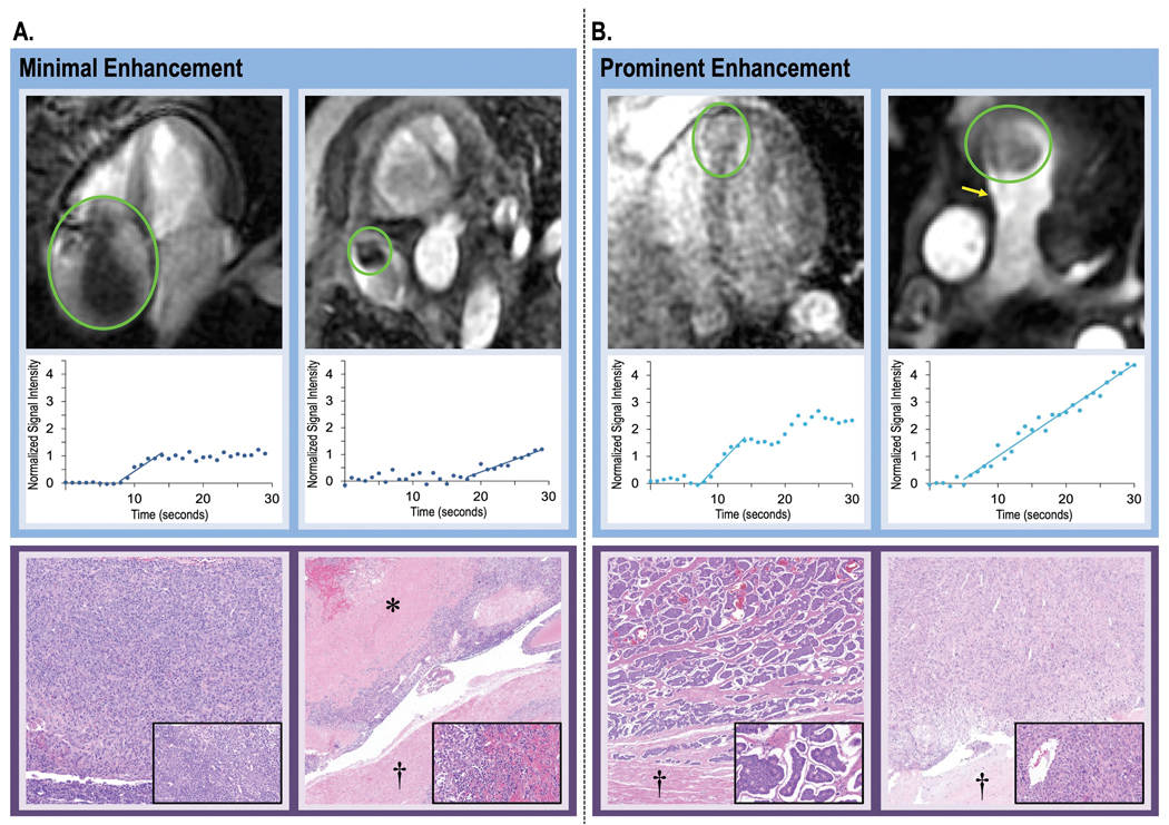
Background: Cardiac magnetic resonance (CMR) differentiates cardiac metastasis (CMET) and cardiac thrombus (CTHR) based on tissue characteristics stemming from vascularity on late gadolinium enhancement (LGE). Perfusion CMR can assess magnitude of vascularity; utility for cardiac masses (CMASS) is unknown.

Objectives: This study sought to determine if perfusion CMR provides diagnostic and prognostic utility for CMASS beyond binary differentiation of CMET and CTHR.

Methods: The population comprised adult cancer patients with CMASS on CMR; CMET and CTHR were defined using LGE-CMR: CMASS+ patients were matched to CMASS- control subjects for cancer type/stage. First-pass perfusion CMR was interpreted visually and semiquantitatively for CMASS vascularity, including contrast enhancement ratio (CER) (plateau vs baseline) and contrast uptake rate (CUR) (slope). Follow-up was performed for all-cause mortality.

Results: A total of 462 cancer patients were studied, including patients with (CMET = 173, CTHR = 69) and without CMASS on LGE-CMR. On perfusion CMR, CER and CUR were higher within CMET vs CTHR (P < 0.001); CUR yielded better performance (AUC: 0.89-0.93) than CER (AUC: 0.66-0.72) (both P < 0.001) to differentiate LGE-CMR-evidenced CMET and CTHR, although both CUR (P = 0.10) and CER (P = 0.01) typically misclassified CMET with minimal enhancement. During follow-up, mortality among CMET patients was high but variable; 47% of patients were alive 1 year post-CMR. Patients with semiquantitative perfusion CMR-evidenced CMET had higher mortality than control subjects (HR: 1.42 [95% CI: 1.06-1.90]; P = 0.02), paralleling visual perfusion CMR (HR: 1.47 [95% CI: 1.12-1.94]; P = 0.006) and LGE-CMR (HR: 1.52 [95% CI: 1.16-2.00]; P = 0.003). Among patients with CMET on LGE-CMR, mortality was highest among patients (P = 0.002) with lesions in the bottom perfusion (CER) tertile, corresponding to low vascularity. Among CMET and cancer-matched control subjects, mortality was equivalent (P = NS) among patients with lesions in the upper CER tertile (corresponding to higher lesion vascularity). Conversely, patients with CMET in the middle (P = 0.03) and lowest (lowest vascularity) (P = 0.001) CER tertiles had increased mortality.

Conclusions: Perfusion CMR yields prognostic utility that complements LGE-CMR: Among cancer patients with LGE-CMR defined CMET, mortality increases in proportion to magnitude of lesion hypoperfusion.

Keywords: cardiac magnetic resonance; cardiac masses; cardio-oncology; late gadolinium enhancement; perfusion.


Conflict of interest statement

Funding Support and Author Disclosures This work was supported by the National Institutes of Health 1R01HL151686 (to Dr Weinsaft), AHA18CDA34080090 and NYS DOH01-ROWLY6 2021- 00048 (to Dr Chan), and MSK SKI Core Grant P30 CA008748. Dr Chan holds a secondary appointment in the Mount Sinai Department of Pharmacology for basic science research unrelated to this study. Dr Weinsaft has received speaker fees from GE Healthcare for talks on cardiac magnetic resonance imaging; and serves as a consultant for Bitterroot Bio. All other authors have reported that they have no relationships relevant to the contents of this paper to disclose.

Figures

Figure 1.
Figure 1.. Representative Examples
Representative examples of left (1A) and right (1B) sided CMET and CTHR as respectively assessed by LGE and perfusion-CMR (top), as well as corresponding perfusion analyses to measure contrast kinetics (bottom). In brief, regions of interest (ROI) were placed over lesions and tracked throughout the cardiac cycle so as to measure slope (contrast uptake rate [CUR]) and plateau enhancement (contrast enhancement ratio [CER]). Note that for both examples, CMET (blue) demonstrated higher contrast enhancement than did CTHR (purple), consistent with presence of vascular supply. However, a range of vascularity was evident among CMET cases for which lesions with minimal enhancement on LGE-CMR (center; dark blue outline) demonstrated lower magnitude of semi-quantitative perfusion than did diffusely enhancing lesions (left; light blue outline), corresponding to lesser differentiation from CTHR.
Figure 2.
Figure 2.. Perfusion-CMR for Differentiation of Cardiac Metastasis and Thrombus
Receiver operating characteristics (ROC) curves for perfusion-CMR cutoffs in relation to CMET as established by the reference of LGE-CMR (red = contrast uptake rate [CUR], blue = enhancement ratio [CER]; solid and dashed lines correspond to absolute and blood pool normalized data for each). Note good overall diagnostic performance for semi-quantitative CUR (AUC 0.89–0.93), which was higher than that of CER (0.66–0.72). Corresponding diagnostic test parameters derived from ROC curves shown in Table 3.
Figure 3.
Figure 3.. CMET Perfusion on CMR in Relation to Pathology-Evidenced Lesion Vascularity
Representative examples of CMET with variable magnitude of perfusion (top) together with semi-quantitative perfusion-CMR analysis (middle) and pathology-evidenced lesion vascularity (bottom). As shown, variability in contrast kinetics on perfusion-CMR paralleled differential vascular and cell density on pathology. 3A: Two cases of minimally enhancing CMET on perfusion-CMR, including thymoma (left) for which pathology demonstrated high tumor cell burden with minimal vascular content and melanoma (right) for which pathology demonstrated scattered vascular content (melanoma) with prominent tumor necrosis. 3B: Two cases of prominently enhancing CMET on perfusion-CMR (left: neuroendocrine, right: spindle cell sarcoma), both of which had high vascular density on pathology. Legend: asterisk = tumor necrosis, dagger = myocardium; inserts = high power views of tumor components (pathology images obtained using hematoxylin and eosin staining).
Figure 4.
Figure 4.. Mortality Based on Diagnostic Categorization by CMR Tissue Characterization Methods
Kaplan-Meier analyses for patients with CMET and CTHR as established by LGE (left), semi-quantitative perfusion-CMR (middle) and visual perfusion-CMR (right), as well as among respective cancer-matched controls. As shown, both CMR tissue characterization methods demonstrated mortality to be higher among patients with CMET, although adverse prognosis conferred by LGE was higher (HR=1.52 [1.16–2.00], p=0.003) than that conferred by perfusion, irrespective of whether interpreted using semi-quantitative (HR=1.42 [1.06–1.90], p=0.02) or visual (HR=1.47 [1.12–1.94], p=0.006) based diagnostic classifications. Conversely, LGE-CMR demonstrated patients with CTHR to have similar mortality to controls, whereas mortality was higher when CTHR was established based on perfusion, consistent with study analysis showing tumors with minimal enhancement (on LGE) to often be misclassified as CTHR based on perfusion cutoffs.
Figure 5.
Figure 5.. Mortality Among Patients with Cardiac Metastases in Relation to Lesion Hypoperfusion
5A: Kaplan-Meier curve encompassing patients with CMET (defined by the reference of LGE-CMR) stratified based on population-based tertiles of lesion vascularity (CER) as measured on semi-quantitative perfusion-CMR. As shown, there was a stepwise association between CMET hypoperfusion and risk for death: Patients in the lowest tertile had higher mortality (p=0.002) than those in the upper-most tertile, paralleling a similar trend for patients in the middle tertile (p=0.09). Primary data from which tertile partitions derived shown in upper right. 5B: Comparisons between CMET cases and cancer-matched controls within each respective semi-quantitative perfusion tertile. As shown, patients within lowest tertile of perfusion-CMR evidenced vascularity (CER) had higher risk for death than did controls (p=0.001), paralleling a similar pattern but lesser magnitude of difference in the middle perfusion tertile (p=0.03). Conversely, mortality for patients with lesions in the highest perfusion tertile (corresponding to highest vascularity) was equivalent to that of cancer-matched controls (p=0.67).
Central Illustration
Central Illustration. Perfusion CMR for Cancer-Associated Cardiac Masses
Multicenter data from two cancer centers demonstrated perfusion-CMR evidenced contrast kinetics to differ between vascular and avascular masses, as evidenced by higher uptake (slope) and plateau enhancement for cardiac metastasis vs. thrombus. While perfusion performed well in relation to LGE, performance as a primary method to differentiate mass types and stratify prognosis was compromised by misclassification of metastases with minimal enhancement: Among patients with CMET on LGE-CMR, perfusion-CMR demonstrated mortality to increase with lesion hypo-perfusion and to be augmented for patients with low vascularity lesions and equivalent (to that of controls) for patients with high vascularity lesions. Findings support utility of integrated CMR tissue characterization for cardiac masses, in which initial diagnosis is established by LGE and prognosis for CMET further stratified based on perfusion-CMR evidenced lesion hypo-vascularity.

Comment in

Similar articles

Cited by

References

    1. Sung H, Ferlay J, Siegel RL et al. Global Cancer Statistics 2020: GLOBOCAN Estimates of Incidence and Mortality Worldwide for 36 Cancers in 185 Countries. CA Cancer J Clin 2021;71:209–249. - PubMed
    1. Weinsaft JW, Kim HW, Shah DJ et al. Detection of left ventricular thrombus by delayed-enhancement cardiovascular magnetic resonance prevalence and markers in patients with systolic dysfunction. J Am Coll Cardiol 2008;52:148–57. - PubMed
    1. Weinsaft JW, Kim HW, Crowley AL et al. LV thrombus detection by routine echocardiography: insights into performance characteristics using delayed enhancement CMR. JACC Cardiovasc Imaging 2011;4:702–12. - PMC - PubMed
    1. Chan AT, Dinsfriend W, Kim J et al. Risk stratification of cardiac metastases using late gadolinium enhancement cardiovascular magnetic resonance: prognostic impact of hypo-enhancement evidenced tumor avascularity. J Cardiovasc Magn Reson 2021;23:42. - PMC - PubMed
    1. Chan AT, Plodkowski AJ, Pun SC et al. Prognostic utility of differential tissue characterization of cardiac neoplasm and thrombus via late gadolinium enhancement cardiovascular magnetic resonance among patients with advanced systemic cancer. J Cardiovasc Magn Reson 2017;19:76. - PMC - PubMed

Publication types

LinkOut - more resources