

CD63/81 Small Extracellular Vesicles in the Aqueous Humor are Retinoblastoma Associated
- PMID: 37410475
- PMCID: PMC10337798
- DOI: 10.1167/iovs.64.10.5
CD63/81 Small Extracellular Vesicles in the Aqueous Humor are Retinoblastoma Associated
Abstract
Purpose: Although biopsy is contraindicated in retinoblastoma (RB), the aqueous humor (AH) is a robust liquid biopsy source of molecular tumor information, facilitating biomarker discovery. Small extracellular vesicles (sEVs), promising biomarker candidates across multiple cancers, were recently identified in RB AH, but relationships between sEVs and RB clinical features are unknown.
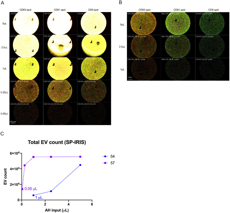
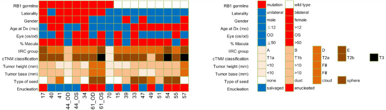
Methods: We analyzed sEVs in 37 AH samples from 18 RB eyes of varying International Intraocular Retinoblastoma Classification (IIRC) groups and explored clinical correlations. Ten samples were collected at diagnosis (DX) and 27 during treatment (Tx). Unprocessed AH underwent Single Particle-Interferometric Reflectance Imaging Sensor (SP-IRIS) analysis for fluorescent particle count and tetraspanin immunophenotyping; counts were subsequentially converted to percentages for analysis.
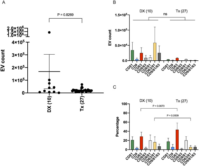
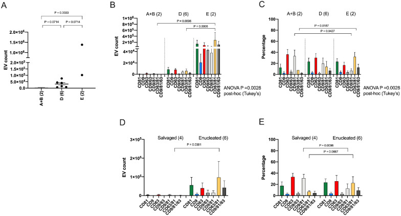
Results: Comparing DX and Tx samples, a higher percentage of CD63/81+ sEVs was found in DX AH (16.3 ± 11.6% vs. 5.49 ± 3.67% P = 0.0009), with a more homogenous mono-CD63+ sEV population seen in Tx AH (43.5 ± 14.7% vs. 28.8 ± 9.38%, P = 0.0073). Among DX samples, CD63/81+ sEVs were most abundant in group E eyes (n = 2) compared to group D (n = 6) by count (2.75 × 10⁵ ± 3.40 × 10⁵ vs. 5.95 × 10³ ± 8.16 × 10³, P = 0.0006), and to group A + B (n = 2) by count (2.75 × 10⁵ ± 3.40 × 10⁵ vs. 2.73 × 10² ± 2.59 × 10², P = 0.0096) and percentage (32.1 ± 7.98% vs. 7.79 ± 0.02%, P = 0.0187).
Conclusions: CD63/81+ sEVs enrich AH from RB eyes before treatment and those with more significant tumor burden, suggesting they are tumor-derived. Future research into their cargo may reveal mechanisms of cellular communication via sEVs in RB and novel biomarkers.
Conflict of interest statement
Disclosure:
Figures
Similar articles
-
Comparative Single Vesicle Analysis of Aqueous Humor Extracellular Vesicles before and after Radiation in Uveal Melanoma Eyes.Int J Mol Sci. 2024 May 30;25(11):6035. doi: 10.3390/ijms25116035. Int J Mol Sci. 2024. PMID: 38892225 Free PMC article.
-
Phenotypic Biomarkers of Aqueous Extracellular Vesicles from Retinoblastoma Eyes.Int J Mol Sci. 2024 Oct 30;25(21):11660. doi: 10.3390/ijms252111660. Int J Mol Sci. 2024. PMID: 39519212 Free PMC article.
-
Single vesicle analysis of aqueous humor in pediatric ocular diseases reveals eye specific CD63-dominant subpopulations.J Extracell Biol. 2022 Apr;1(4):e36. doi: 10.1002/jex2.36. Epub 2022 Apr 20. J Extracell Biol. 2022. PMID: 36339649 Free PMC article.
-
The Potential of Aqueous Humor Sampling in Diagnosis, Prognosis, and Treatment of Retinoblastoma.Invest Ophthalmol Vis Sci. 2024 Jan 2;65(1):18. doi: 10.1167/iovs.65.1.18. Invest Ophthalmol Vis Sci. 2024. PMID: 38180770 Free PMC article. Review.
-
Aqueous Humor Markers in Retinoblastoma, a Review.Transl Vis Sci Technol. 2019 Apr 9;8(2):13. doi: 10.1167/tvst.8.2.13. eCollection 2019 Apr. Transl Vis Sci Technol. 2019. PMID: 31019846 Free PMC article. Review.
Cited by
-
An Eye on Extracellular Vesicles: Trends and Clinical Translations in Vision Research.Ophthalmol Sci. 2024 Sep 12;5(1):100619. doi: 10.1016/j.xops.2024.100619. eCollection 2025 Jan-Feb. Ophthalmol Sci. 2024. PMID: 39584184 Free PMC article.
-
Comparative Single Vesicle Analysis of Aqueous Humor Extracellular Vesicles before and after Radiation in Uveal Melanoma Eyes.Int J Mol Sci. 2024 May 30;25(11):6035. doi: 10.3390/ijms25116035. Int J Mol Sci. 2024. PMID: 38892225 Free PMC article.
-
Phenotypic Biomarkers of Aqueous Extracellular Vesicles from Retinoblastoma Eyes.Int J Mol Sci. 2024 Oct 30;25(21):11660. doi: 10.3390/ijms252111660. Int J Mol Sci. 2024. PMID: 39519212 Free PMC article.
-
Retinoblastoma: Aqueous humor liquid biopsy.Taiwan J Ophthalmol. 2025 Mar 20;15(1):55-61. doi: 10.4103/tjo.TJO-D-24-00133. eCollection 2025 Jan-Mar. Taiwan J Ophthalmol. 2025. PMID: 40213298 Free PMC article. Review.
-
Advances in Extracellular-Vesicles-Based Diagnostic and Therapeutic Approaches for Ocular Diseases.ACS Nano. 2024 Aug 27;18(34):22793-22828. doi: 10.1021/acsnano.4c08486. Epub 2024 Aug 14. ACS Nano. 2024. PMID: 39141830 Free PMC article. Review.
References
-
- Roy SR, Kaliki S. Retinoblastoma: A MAJOR REVIEW. Mymensingh Med J. 2021; 30(3): 881–895. - PubMed
-
- Shields JA, Shields CL, Ehya H, Eagle RC, De Potter P.. Fine-needle aspiration biopsy of suspected intraocular tumors. Int Ophthalmol Clin. 1993; 33(3): 77–82. - PubMed
-
- Karcioglu ZA. Fine needle aspiration biopsy (FNAB) for retinoblastoma. Retina. 2002; 22(6): 707–710. - PubMed
-
- Karcioglu ZA, Gordon RA, Karcioglu GL.. Tumor seeding in ocular fine needle aspiration biopsy. Ophthalmology. 1985; 92(12): 1763–1767. - PubMed
-
- Eide N, Syrdalen P, Walaas L, Hagmar B.. Fine needle aspiration biopsy in selecting treatment for inconclusive intraocular disease. Acta Ophthalmol Scand. 1999; 77(4): 448–452. - PubMed
MeSH terms
Substances
Grants and funding
LinkOut - more resources
Full Text Sources
Miscellaneous
