. 2023 Jul 18;120(29):e2304870120.
doi: 10.1073/pnas.2304870120. Epub 2023 Jul 6.

The ubiquitin-protein ligase MIEL1 localizes to peroxisomes to promote seedling oleosin degradation and lipid droplet mobilization

Affiliations

The ubiquitin-protein ligase MIEL1 localizes to peroxisomes to promote seedling oleosin degradation and lipid droplet mobilization

Melissa S Traver et al. Proc Natl Acad Sci U S A. .

Abstract

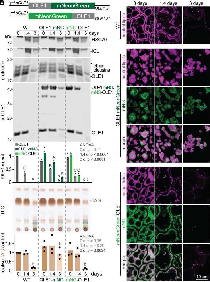
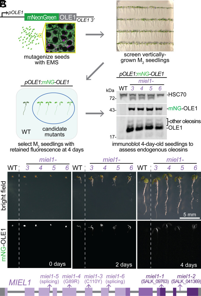
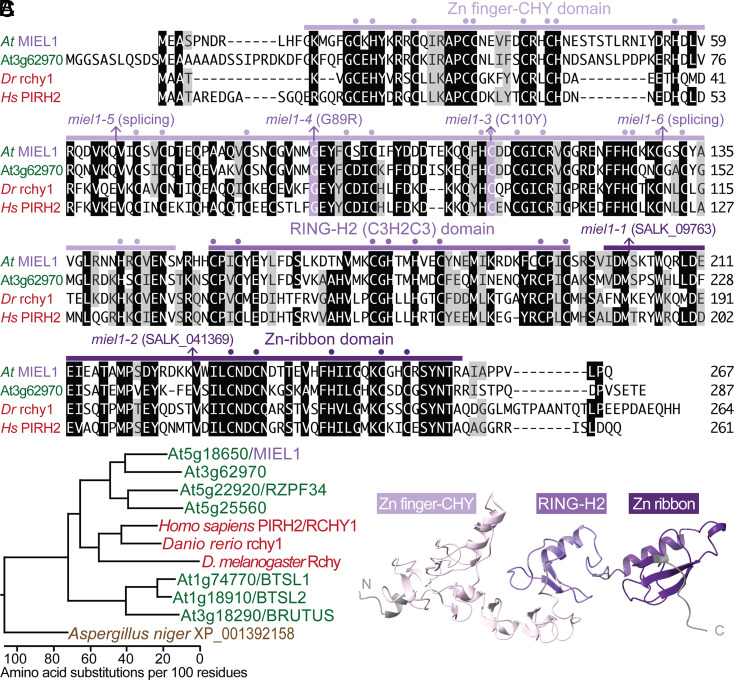
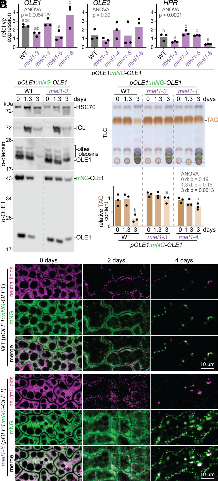
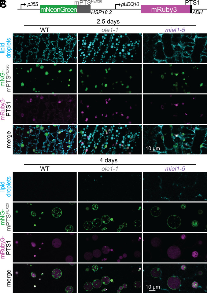
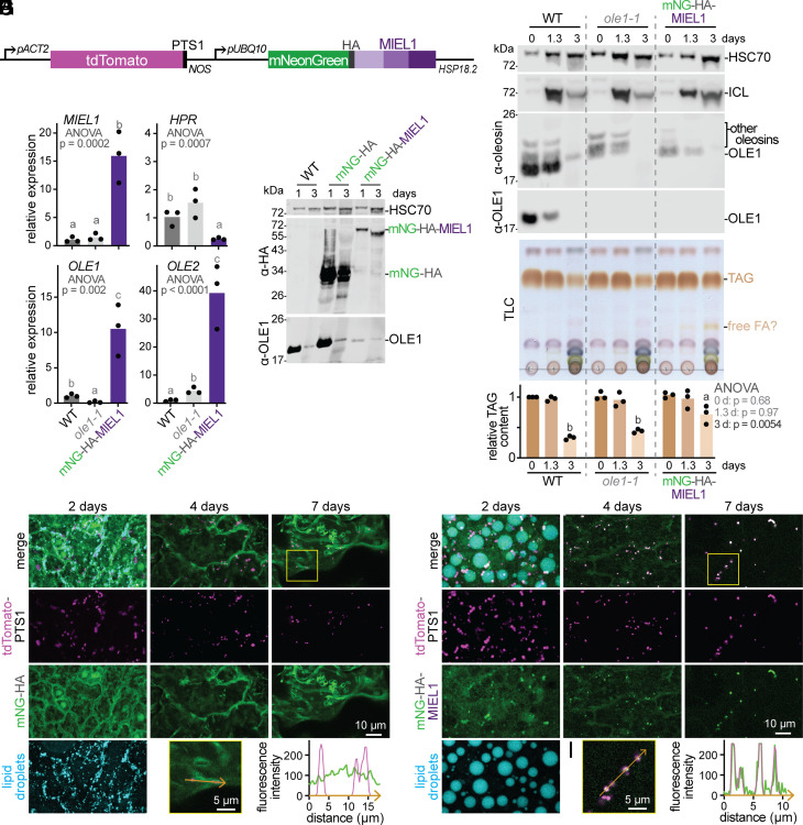
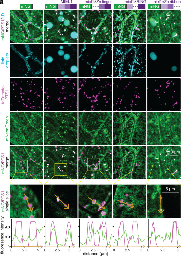
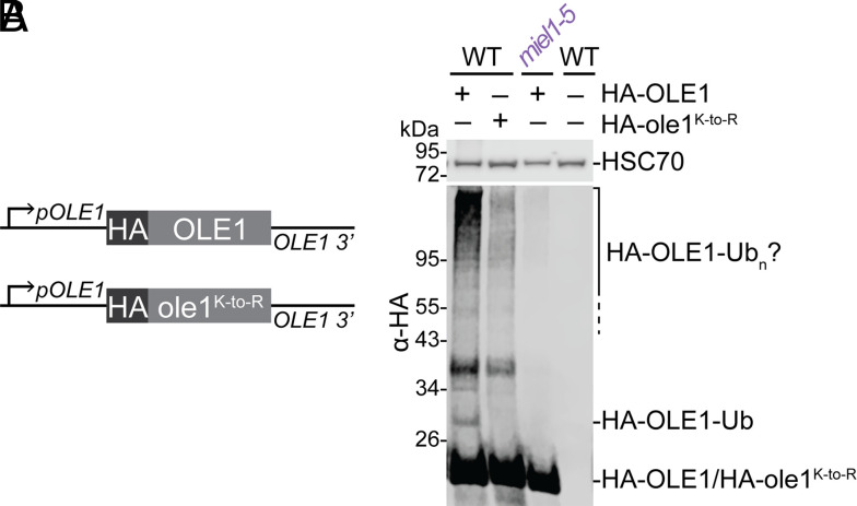
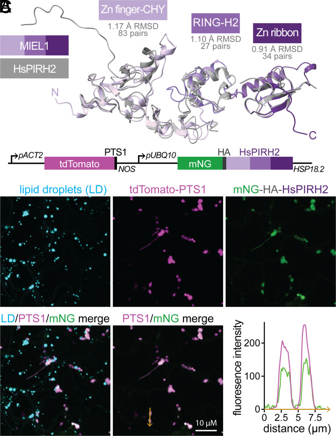
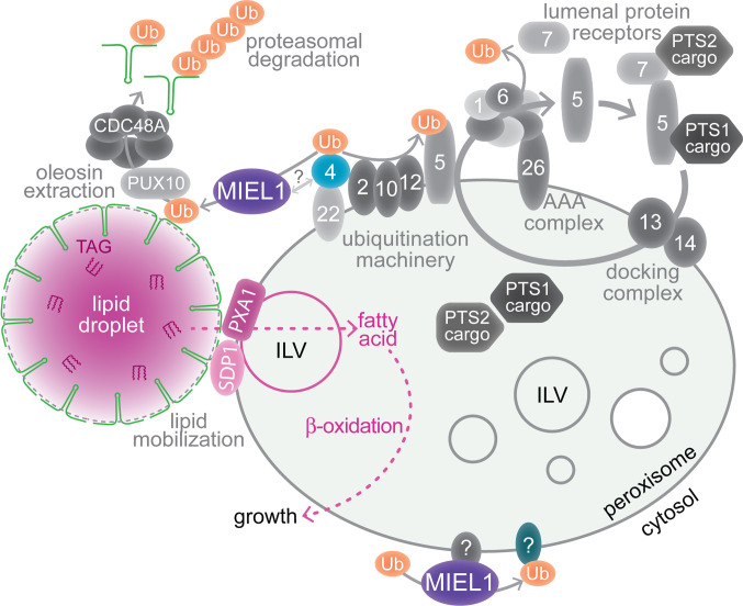
Lipid droplets are organelles conserved across eukaryotes that store and release neutral lipids to regulate energy homeostasis. In oilseed plants, fats stored in seed lipid droplets provide fixed carbon for seedling growth before photosynthesis begins. As fatty acids released from lipid droplet triacylglycerol are catabolized in peroxisomes, lipid droplet coat proteins are ubiquitinated, extracted, and degraded. In Arabidopsis seeds, the predominant lipid droplet coat protein is OLEOSIN1 (OLE1). To identify genes modulating lipid droplet dynamics, we mutagenized a line expressing mNeonGreen-tagged OLE1 expressed from the OLE1 promoter and isolated mutants with delayed oleosin degradation. From this screen, we identified four miel1 mutant alleles. MIEL1 (MYB30-interacting E3 ligase 1) targets specific MYB transcription factors for degradation during hormone and pathogen responses [D. Marino et al., Nat. Commun. 4, 1476 (2013); H. G. Lee and P. J. Seo, Nat. Commun. 7, 12525 (2016)] but had not been implicated in lipid droplet dynamics. OLE1 transcript levels were unchanged in miel1 mutants, indicating that MIEL1 modulates oleosin levels posttranscriptionally. When overexpressed, fluorescently tagged MIEL1 reduced oleosin levels, causing very large lipid droplets. Unexpectedly, fluorescently tagged MIEL1 localized to peroxisomes. Our data suggest that MIEL1 ubiquitinates peroxisome-proximal seed oleosins, targeting them for degradation during seedling lipid mobilization. The human MIEL1 homolog (PIRH2; p53-induced protein with a RING-H2 domain) targets p53 and other proteins for degradation and promotes tumorigenesis [A. Daks et al., Cells 11, 1515 (2022)]. When expressed in Arabidopsis, human PIRH2 also localized to peroxisomes, hinting at a previously unexplored role for PIRH2 in lipid catabolism and peroxisome biology in mammals.

Keywords: Arabidopsis thaliana; E3 ubiquitin ligase; PIRH2; lipid droplet; peroxisome.


Conflict of interest statement

The authors declare no competing interest.

Figures

Fig. 1.
Fig. 1.
A biologically neutral OLE1 reporter. (A) Constructs to express mNeonGreen fused to the OLE1 C (Top) or N (Bottom) terminus flanked by OLE1 5′ and 3′ sequences. (B and C) Seedlings were processed for (B) immunoblotting with the indicated antibodies or (C) TLC of extracted lipids. Graphs indicate relative OLE1 signal relative to HSC70 (B) or TAG content (C) in three biological replicates (including the one shown) normalized to the WT signal at 0 d. Separate one-way ANOVA tests were performed to compare genotypes at each timepoint; letters represent homogenous subsets assigned by Tukey’s posthoc test when the P-value was <0.05. (D) Confocal images (single slices) of cotyledon epidermal cells. Neutral lipids were stained with MDH (magenta); mNeonGreen fluorescence is shown in green.
Fig. 2.
Fig. 2.
Isolation of miel1 mutants in a screen for persistent mNeonGreen–OLE1 fluorescence. (A) pOLE1:mNeonGreen–OLE1 seeds were mutagenized and their progeny was screened for sustained seedling fluorescence following germination. The Lower Right panel shows an immunoblot of 4-d-old unmutagenized pOLE1:mNeonGreen–OLE1 (WT) seedlings and miel1 mutant seedlings. (B) Representative bright field (Top) and mNeonGreen–OLE1 fluorescence (Bottom, grayscale) images of WT and miel1 mutants during germination and seedling development. (C) Gene diagram of MIEL1 showing newly identified (miel1-3 to miel1-6) and previously described (miel1-1 and miel1-2) mutations. Exons (boxes) are connected by introns (lines). Shades of purple denote MIEL1 structural domains depicted in Fig. 3.
Fig. 3.
Fig. 3.
MIEL1 is a conserved Cys-rich ubiquitin-protein ligase. (A) Alignment of MIEL1 and related proteins generated using the MegAlign (DNAStar) Clustal W method (BLOSUM series protein weight matrix). Identical residues are highlighted in black; chemically similar residues are highlighted in gray. miel1 mutations, conserved Cys and His residues, and MIEL1 domains are indicated above the sequences. (B) Phylogenetic tree including additional MIEL1 relatives constructed using the MegAlign program from the alignment shown in SI Appendix, Fig. S1. (C) Arabidopsis MIEL1 structure predicted by AlphaFold (41, 42) depicting the Zn finger-CHY (light purple), RING-H2 (medium purple), and Zn ribbon (dark purple) domains.
Fig. 4.
Fig. 4.
miel1 mutants retain oleosins, TAG, and lipid droplets longer than wild-type seedlings but do not display elevated oleosin transcript levels. (A) Relative OLE1,OLE2, and HPR transcript levels in 2-d-old seedlings measured via RT-qPCR. Letters above bars represent homogenous subsets assigned by Tukey’s posthoc test when one-way ANOVA P-values were <0.05. (B) Immunoblot probed with the indicated antibodies. Molecular weight marker positions (in kDa) are at the left. (C) TLC of extracted lipids. The graph shows relative TAG content in three biological replicates (including the one shown) normalized to the WT signal at 0 d and statistically analyzed as in the legend of Fig. 1. (D) Confocal images (single slices) of WT and miel1-6 cotyledon epidermal cells. Neutral lipids were stained with MDH (magenta); mNeonGreen–OLE1 fluorescence is shown in green. In panels BD, 0-day samples were stratified seeds; other samples were seedlings harvested at the indicated times after sowing.
Fig. 5.
Fig. 5.
MIEL1 loss appears to decrease lipid droplet size and impact peroxisome morphology. (A) Diagram of constitutively expressed fluorescent reporters marking peroxisomal membranes (mNeonGreen–mPTSPEX26) and lumen (mRuby3–PTS1) (39). (B and C) Confocal images (single slices) of MDH-stained neutral lipids (cyan), peroxisome membranes (green), and peroxisome lumen (magenta) in cotyledon epidermal cells of 2.5- (B) and 4- (C) d-old WT, ole1-1, and miel1-5 seedlings expressing the reporters diagramed in panel A.
Fig. 6.
Fig. 6.
Overexpressed mNeonGreen–MIEL1 decreases oleosin levels, slows TAG utilization, enlarges lipid droplets, and localizes to peroxisomes. (A) Diagram of constitutively expressed fluorescent reporters marking peroxisomal lumen (tdTomato–PTS1) and MIEL1 (mNeonGreen–HA–MIEL1). (B) Relative mRNA levels in 2-d-old seedlings were measured via RT-qPCR and statistically analyzed as in the legend of Fig. 4. (C) Immunoblot of seedlings without a reporter (WT), segregating for mNeonGreen–HA, or homozygous for mNeonGreen–HA–MIEL1 probed with the indicated antibodies. (D) Immunoblot probed with the indicated antibodies. (E) TLC of extracted lipids. The graph shows relative TAG content in three biological replicates (including the one shown) normalized to the WT signal at 0 d and statistically analyzed as in the legend of Fig. 1. (F) Confocal images (maximum-intensity projections of twelve 1-µm slices) of hypocotyl epidermal cells showing tdTomato–PTS1 (magenta) and mNeonGreen–HA (green) at the indicated seedling ages. Neutral lipids were stained with MDH (cyan) in 2-d-old seedlings. (G) Magnification of boxed region in 7-d image from panel F. Plot shows tdTomato–PTS1 (magenta) and mNeonGreen–HA (green) fluorescence intensity across multiple peroxisomes (orange arrow). (H) As in panel F, but with mNeonGreen–HA–MIEL1. (I) Magnification of boxed region in 7-d image from panel H. Plot shows tdTomato–PTS1 (magenta) and mNeonGreen–HA–MIEL1 (green) fluorescence intensity across multiple peroxisomes (orange arrow). In panels CE, 0-d samples were stratified seeds; other samples were seedlings harvested at the indicated times following sowing.
Fig. 7.
Fig. 7.
The MIEL1 N-terminal and RING domains direct peroxisome localization and lipid droplet enlargement. (A) Lipid droplets (stained with MDH; cyan); peroxisomes (tdTomato–PTS1; magenta); and either mNeonGreen–HA, mNeonGreen–HA–MIEL1, or mNeonGreen–HA–MIEL1 domain deletion derivatives (green) were visualized in hypocotyl epidermal cells of 2-d-old seedlings. Maximum-intensity projections of ten 1-µm slices are shown. (B) Magnification of single slices from the boxed regions in panel A. Plots show tdTomato–PTS1 (magenta) and mNeonGreen (green) fluorescence intensity across multiple peroxisomes (orange arrows).
Fig. 8.
Fig. 8.
MIEL1 is necessary for OLE1 ubiquitination. (A) Constructs to express HA-tagged OLE1 or ole1K-toR (with Lys residues altered to Arg) flanked by OLE1 5′ and 3′ sequences. (B) Immunoblot of proteins from 1-d-old WT seedlings expressing HA–OLE1 or HA–ole1K-toR, miel1-5 expressing HA–OLE1, or untransformed WT.
Fig. 9.
Fig. 9.
The human MIEL1 homolog HsPIRH2 localizes to peroxisomes when expressed in Arabidopsis. (A) HsPIRH2 NMR structures (gray) (PDB 2K2C, 2JRJ, and 2K2D) (51) were aligned to the MIEL1 structure (purple) predicted by AlphaFold (41, 42). (B) Diagram of constitutively expressed fluorescent reporters marking peroxisomal lumen (tdTomato–PTS1) and human PIRH2 (mNeonGreen–HA–HsPIRH2). (C) Cotyledon epidermal cells of 4-d-old seedlings transformed with the construct shown in panel B were imaged using confocal microscopy. Maximum-intensity projections (ten 1-µm slices) of neutral lipids stained with MDH (cyan), tdTomato–PTS1 (magenta), and mNeonGreen–HA–HsPIRH2 (green) are shown. The plot shows tdTomato–PTS1 (magenta) and mNeonGreen (green) fluorescence intensity across two peroxisomes (orange arrow).
Fig. 10.
Fig. 10.
Working model for MIEL1 functions at peroxisomes. MIEL1 is necessary for oleosin (green hairpins) ubiquitination and timely degradation during lipid mobilization. Oleosin ubiquitination allows extraction by PUX10 and CDC48A (20, 21) and proteasomal degradation. MIEL1 localizes at peroxisomes, perhaps via interaction with an organelle surface protein, such as the ubiquitin-conjugating enzyme PEX4. PEX4 (blue) and other peroxins (numbered shapes) function in the import of lumenal cargo proteins with PTS1 or PTS2 signals via the PEX7 and PEX7 cycling receptors. MIEL1 may also target proteins on the peroxisome that modulate peroxisome dynamics, such as the formation of ILVs, which are implicated in fatty acid β-oxidation and protein sorting (39). SDP1 is a peroxisome-associated TAG lipase (34, 35), and PXA1 transports fatty acids into the organelle for β-oxidation (36, 37).

Similar articles

Cited by

References

    1. Huang A. H. C., Plant lipid droplets and their associated proteins: Potential for rapid advances. Plant Physiol. 176, 1894–1918 (2018). - PMC - PubMed
    1. Lundquist P. K., Shivaiah K.-K., Espinoza-Corral R., Lipid droplets throughout the evolutionary tree. Prog. Lipid Res. 78, 101029 (2020). - PubMed
    1. Guzha A., Whitehead P., Ischebeck T., Chapman K. D., Lipid droplets: packing hydrophobic molecules within the aqueous cytoplasm. Annu. Rev. Plant Biol. 74, 195–223 (2023). - PubMed
    1. Listenberger L. L., et al. , Triglyceride accumulation protects against fatty acid-induced lipotoxicity. Proc. Natl. Acad. Sci. U.S.A. 100, 3077–3082 (2003). - PMC - PubMed
    1. Ischebeck T., Krawczyk H. E., Mullen R. T., Dyer J. M., Chapman K. D., Lipid droplets in plants and algae: Distribution, formation, turnover and function. Semin. Cell Dev. Biol. 108, 82–93 (2020). - PubMed

MeSH terms