

Transactive response DNA-binding protein 43 is enriched at the centrosome in human cells
- PMID: 37410912
- PMCID: PMC10473568
- DOI: 10.1093/brain/awad228
Transactive response DNA-binding protein 43 is enriched at the centrosome in human cells
Abstract
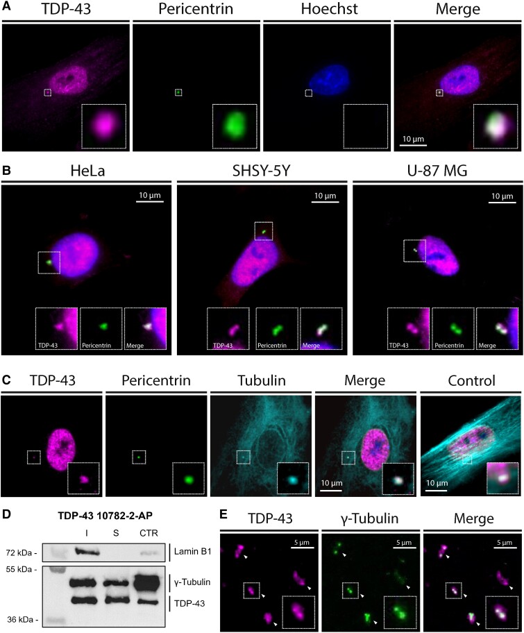
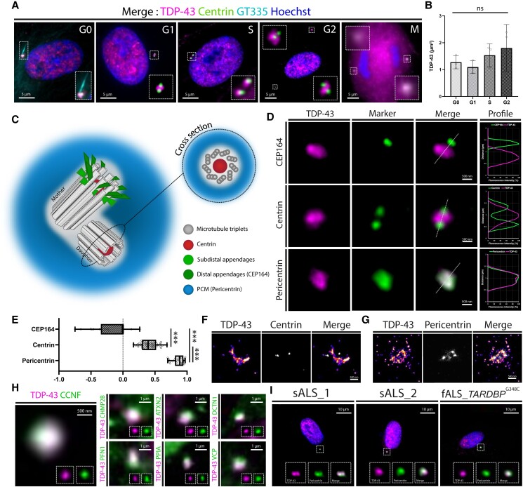
The centrosome, as the main microtubule organizing centre, plays key roles in cell polarity, genome stability and ciliogenesis. The recent identification of ribosomes, RNA-binding proteins and transcripts at the centrosome suggests local protein synthesis. In this context, we hypothesized that TDP-43, a highly conserved RNA binding protein involved in the pathophysiology of amyotrophic lateral sclerosis and frontotemporal lobar degeneration, could be enriched at this organelle. Using dedicated high magnification sub-diffraction microscopy on human cells, we discovered a novel localization of TDP-43 at the centrosome during all phases of the cell cycle. These results were confirmed on purified centrosomes by western blot and immunofluorescence microscopy. In addition, the co-localization of TDP-43 and pericentrin suggested a pericentriolar enrichment of the protein, leading us to hypothesize that TDP-43 might interact with local mRNAs and proteins. Supporting this hypothesis, we found four conserved centrosomal mRNAs and 16 centrosomal proteins identified as direct TDP-43 interactors. More strikingly, all the 16 proteins are implicated in the pathophysiology of TDP-43 proteinopathies, suggesting that TDP-43 dysfunction in this organelle contributes to neurodegeneration. This first description of TDP-43 centrosomal enrichment paves the way for a more comprehensive understanding of TDP-43 physiology and pathology.
Keywords: ALS; FTLD; TDP-43; centrosome; pericentriolar matrix.
© The Author(s) 2023. Published by Oxford University Press on behalf of the Guarantors of Brain.
Conflict of interest statement
The authors report no competing interest.
Figures
Comment in
-
The MLO-down on TDP-43.Brain. 2023 Sep 1;146(9):3565-3567. doi: 10.1093/brain/awad268. Brain. 2023. PMID: 37540028 Free PMC article.
Similar articles
-
[FTLD/ALS as TDP-43 proteinopathies].Rinsho Shinkeigaku. 2010 Nov;50(11):1022-4. doi: 10.5692/clinicalneurol.50.1022. Rinsho Shinkeigaku. 2010. PMID: 21921552 Review. Japanese.
-
Mislocalization of Nup62 Contributes to TDP-43 Proteinopathy in ALS/FTLD.ACS Chem Neurosci. 2022 Sep 7;13(17):2544-2546. doi: 10.1021/acschemneuro.2c00480. Epub 2022 Aug 24. ACS Chem Neurosci. 2022. PMID: 36001801
-
Disease animal models of TDP-43 proteinopathy and their pre-clinical applications.Int J Mol Sci. 2013 Oct 9;14(10):20079-111. doi: 10.3390/ijms141020079. Int J Mol Sci. 2013. PMID: 24113586 Free PMC article. Review.
-
TDP-43 aggregation in neurodegeneration: are stress granules the key?Brain Res. 2012 Jun 26;1462:16-25. doi: 10.1016/j.brainres.2012.02.032. Epub 2012 Feb 22. Brain Res. 2012. PMID: 22405725 Free PMC article. Review.
-
Physiological functions and pathobiology of TDP-43 and FUS/TLS proteins.J Neurochem. 2016 Aug;138 Suppl 1:95-111. doi: 10.1111/jnc.13625. Epub 2016 Jun 15. J Neurochem. 2016. PMID: 27015757 Review.
Cited by
-
Amyotrophic lateral sclerosis and frontotemporal dementia mutation reduces endothelial TDP-43 and causes blood-brain barrier defects.Sci Adv. 2025 Apr 18;11(16):eads0505. doi: 10.1126/sciadv.ads0505. Epub 2025 Apr 16. Sci Adv. 2025. PMID: 40238886 Free PMC article.
-
Loss of Endothelial TDP-43 Leads to Blood Brain Barrier Defects in Mouse Models of Amyotrophic Lateral Sclerosis and Frontotemporal Dementia.bioRxiv [Preprint]. 2023 Dec 14:2023.12.13.571184. doi: 10.1101/2023.12.13.571184. bioRxiv. 2023. Update in: Sci Adv. 2025 Apr 18;11(16):eads0505. doi: 10.1126/sciadv.ads0505. PMID: 38168388 Free PMC article. Updated. Preprint.
-
Centrosomes and cilia in neurodegeneration: main actors or mere spectators?Open Biol. 2025 May;15(5):240317. doi: 10.1098/rsob.240317. Epub 2025 May 21. Open Biol. 2025. PMID: 40393509 Free PMC article. Review.
-
The MLO-down on TDP-43.Brain. 2023 Sep 1;146(9):3565-3567. doi: 10.1093/brain/awad268. Brain. 2023. PMID: 37540028 Free PMC article.
-
Colchicine treatment in amyotrophic lateral sclerosis: safety, biological and clinical effects in a randomized clinical trial.Brain Commun. 2024 Sep 5;6(5):fcae304. doi: 10.1093/braincomms/fcae304. eCollection 2024. Brain Commun. 2024. PMID: 39291166 Free PMC article. Clinical Trial.
References
-
- Arai T, Hasegawa M, Akiyama H, et al. . TDP-43 is a component of ubiquitin-positive tau-negative inclusions in frontotemporal lobar degeneration and amyotrophic lateral sclerosis. Biochem Biophys Res Commun. 2006;351:602–611. - PubMed
-
- Neumann M, Sampathu DM, Kwong LK, et al. . Ubiquitinated TDP-43 in frontotemporal lobar degeneration and amyotrophic lateral sclerosis. Science. 2006;314:130–133. - PubMed
-
- Kabashi E, Valdmanis PN, Dion P, et al. . TARDBP Mutations in individuals with sporadic and familial amyotrophic lateral sclerosis. Nat Genet. 2008;40:572–574. - PubMed
Publication types
MeSH terms
Substances
LinkOut - more resources
Full Text Sources
Medical
Miscellaneous
