. 2023 Aug;299(8):105014.
doi: 10.1016/j.jbc.2023.105014. Epub 2023 Jul 4.

Heme binding to the SARS-CoV-2 spike glycoprotein

Affiliations

Heme binding to the SARS-CoV-2 spike glycoprotein

Samuel L Freeman et al. J Biol Chem. 2023 Aug.

Abstract

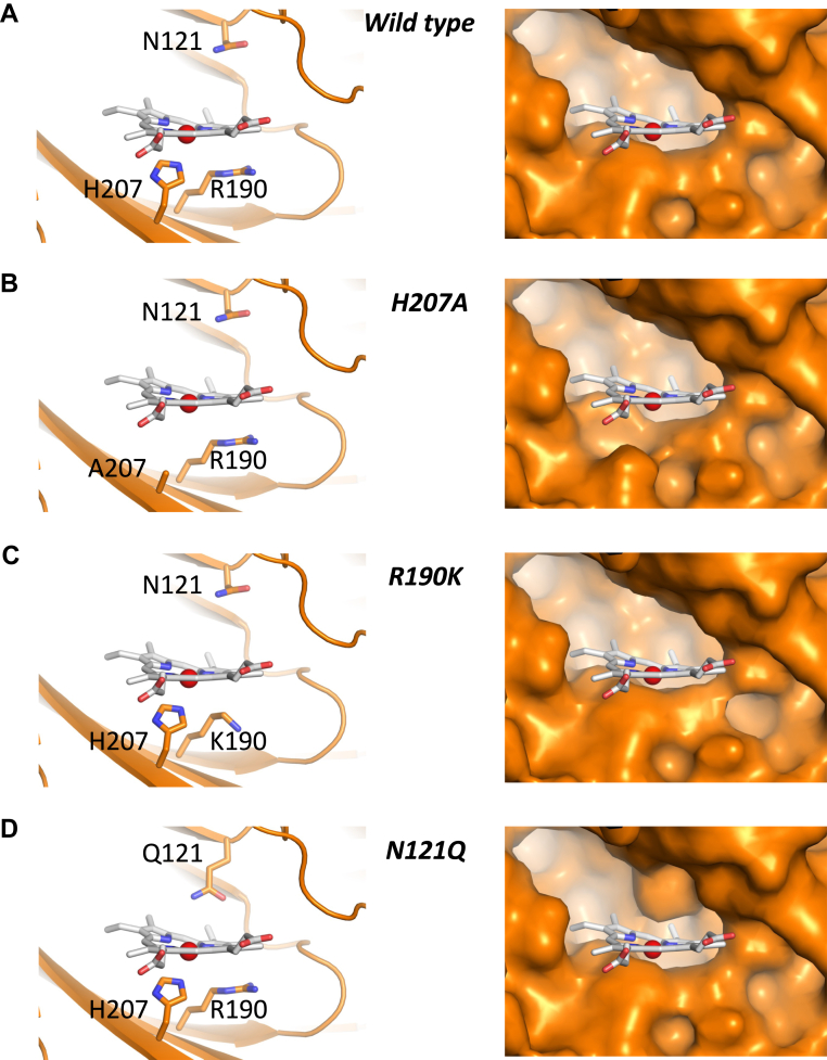
The target for humoral immunity, SARS-CoV-2 spike glycoprotein, has become the focus of vaccine research and development. Previous work demonstrated that the N-terminal domain (NTD) of SARS-CoV-2 spike binds biliverdin-a product of heme catabolism-causing a strong allosteric effect on the activity of a subset of neutralizing antibodies. Herein, we show that the spike glycoprotein is also able to bind heme (KD = 0.5 ± 0.2 μM). Molecular modeling indicated that the heme group fits well within the same pocket on the SARS-CoV-2 spike NTD. Lined by aromatic and hydrophobic residues (W104, V126, I129, F192, F194, I203, and L226), the pocket provides a suitable environment to stabilize the hydrophobic heme. Mutagenesis of N121 has a substantive effect on heme binding (KD = 3000 ± 220 μM), confirming the pocket as a major heme binding location of the viral glycoprotein. Coupled oxidation experiments in the presence of ascorbate indicated that the SARS-CoV-2 glycoprotein can catalyze the slow conversion of heme to biliverdin. The heme trapping and oxidation activities of the spike may allow the virus to reduce levels of free heme during infection to facilitate evasion of the adaptive and innate immunity.

Keywords: Heme; SARS-CoV-2; biliverdin; spike protein.


Conflict of interest statement

Conflicts of interest The authors declare that they have no conflicts of interest with the contents of this article.

Figures

Figure 1
Figure 1
Structures of SARS-CoV-2 spike protein. A, cryo-EM structure (PDB 7NT9) of the trimeric SARS-CoV-2 protein (3RBD-down, in the closed conformation) in complex with the alpha isomer of biliverdin (biliverdin α, in green). The three protomers of the spike protein, forming the trimer, are coloured in blue, orange, and yellow. B, cryo-EM structure (PDB 7NT9) showing an expanded view of the biliverdin binding region of the N-terminal domain. Residues in close proximity to the biliverdin molecule are labeled, and the dashed line from N121 shows an important hydrogen bonding interaction with the tetrapyrrole (pyrrole D).
Figure 2
Figure 2
Spectra of heme-saturated samples of the Wuhan-Hu-1 S1 protein ([heme]:[protein] = 1:1). From top: Wild-type (small dashes), RBD (dots), NTD (dot-dash), H207A (large dashes), R190K (solid grey), and N121Q (solid black). Wavelength maxima for each protein are indicated in Fig. S1. All samples have been normalized at 280 nm for easier comparison.
Figure 3
Figure 3
UV-visible spectra showing the coupled oxidation process over a period of 5 h for the reaction of wild type S1 (2.5 μM) with ascorbate (0.5 mM) under aerobic conditions. Arrows indicate direction of peak movements. Individual spectra were taken at 20-min intervals. After approximately 5 h, no further changes in the spectrum were observed, and the reaction is presumed to have reached completion. LC-MS analysis of the products of this reaction shows evidence of biliverdin formation (observed [M + H]+ = 583.2538 Da (2.1 ppm error), data not shown).
Figure 4
Figure 4
Model of the heme-spike complex. A, surface representation of the heme-binding pocket in the wild type S1. The three protomers are colored as in Figure 1B. Detailed view of the residues lining the proposed heme binding pocket. The green color highlights the location of N121, R190, and H207 (shown in the same orientation as in the left panel of (C)). The heme is colored white with the iron in the center as a red sphere. C, Left: Close up of the heme-binding pocket, with the N121, R190, and H207 residues shown in green, in the same orientation as in (B). Right – The same view as in (A) but with a 30° rotation about the horizontal plane, with other residues relevant to the discussion labeled. The heme pyrrole rings are labeled AD.
Figure 5
Figure 5
Detailed view (left) and space-filling models (right) of the heme-binding pockets. Figure shows heme pockets in (A) wild type, (B) H207A, (C) R190K, and (D) N121Q variants. The N121Q, R190K, and H207A mutations were introduced using Pymol. The heme is colored as in Figure 4. Note that according to the model, the substitution of N121 by a glutamine alters the shape and substantially reduces the total volume contained within the pocket, and thus reduces the space available for heme binding. There is other evidence from in silico analyses for heme binding at this same location (28, 46).
Figure 6
Figure 6
Structural analysis of potential heme binding pockets in pathogenic coronaviruses. (A) Comparison of the proposed heme binding pockets between SARS-CoV-2, SARS-CoV (PDB 5XLR (41)) and MERS-CoV (PDB 5X5C (42)). B, alignments of the heme-bound SARS-CoV-2 spike model (orange) with SARS-CoV (yellow) and MERS-CoV (cyan) structures. The top panel shows the superimposition of the heme-bound SARS-CoV-2 spike model with the cryo-EM SARS-CoV structure. The middle panel displays the superimposition of the heme-bound SARS-CoV-2 spike model with the cryo-EM MERS-CoV structure. The lower panel illustrates the overlapping of all three spike proteins. The heme group and some of the residues lining the heme-binding site are highlighted with sticks.

References

    1. Huang Y., Yang C., Xu X.F., Xu W., Liu S.W. Structural and functional properties of SARS-CoV-2 spike protein: potential antivirus drug development for COVID-19. Acta Pharmacol. Sin. 2020;41:1141–1149. - PMC - PubMed
    1. Lan J., Ge J., Yu J., Shan S., Zhou H., Fan S., et al. Structure of the SARS-CoV-2 spike receptor-binding domain bound to the ACE2 receptor. Nature. 2020;581:215–220. - PubMed
    1. Cerutti G., Guo Y., Wang P., Nair M.S., Huang Y., Yu J., et al. Neutralizing antibody 5-7 defines a distinct site of vulnerability in SARS-CoV-2 spike N-terminal domain. Cell Rep. 2021;37:109928. - PMC - PubMed
    1. Rosa A., Pye V.E., Graham C., Muir L., Seow J., Ng K.W., et al. SARS-CoV-2 can recruit a heme metabolite to evade antibody immunity. Sci. Adv. 2021;7 - PMC - PubMed
    1. Qu K., Chen Q., Ciazynska K.A., Liu B., Zhang X., Wang J., et al. Engineered disulfide reveals structural dynamics of locked SARS-CoV-2 spike. PLoS Pathog. 2022;18 - PMC - PubMed

Publication types