. 2023 Oct:175:116836.
doi: 10.1016/j.bone.2023.116836. Epub 2023 Jul 4.

miR181a/b-1 controls osteocyte metabolism and mechanical properties independently of bone morphology

Affiliations

miR181a/b-1 controls osteocyte metabolism and mechanical properties independently of bone morphology

Jihee Yoon et al. Bone. 2023 Oct.

Abstract

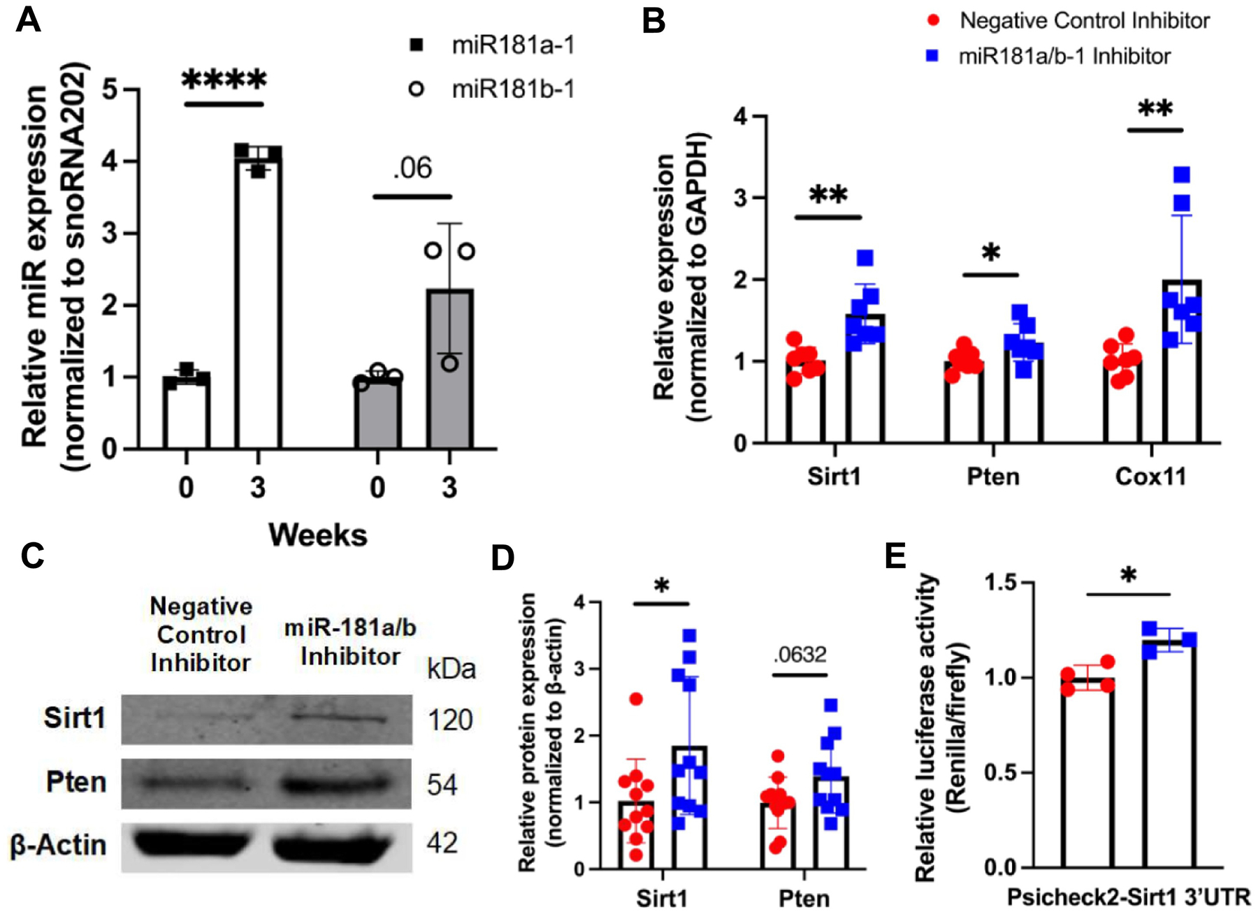
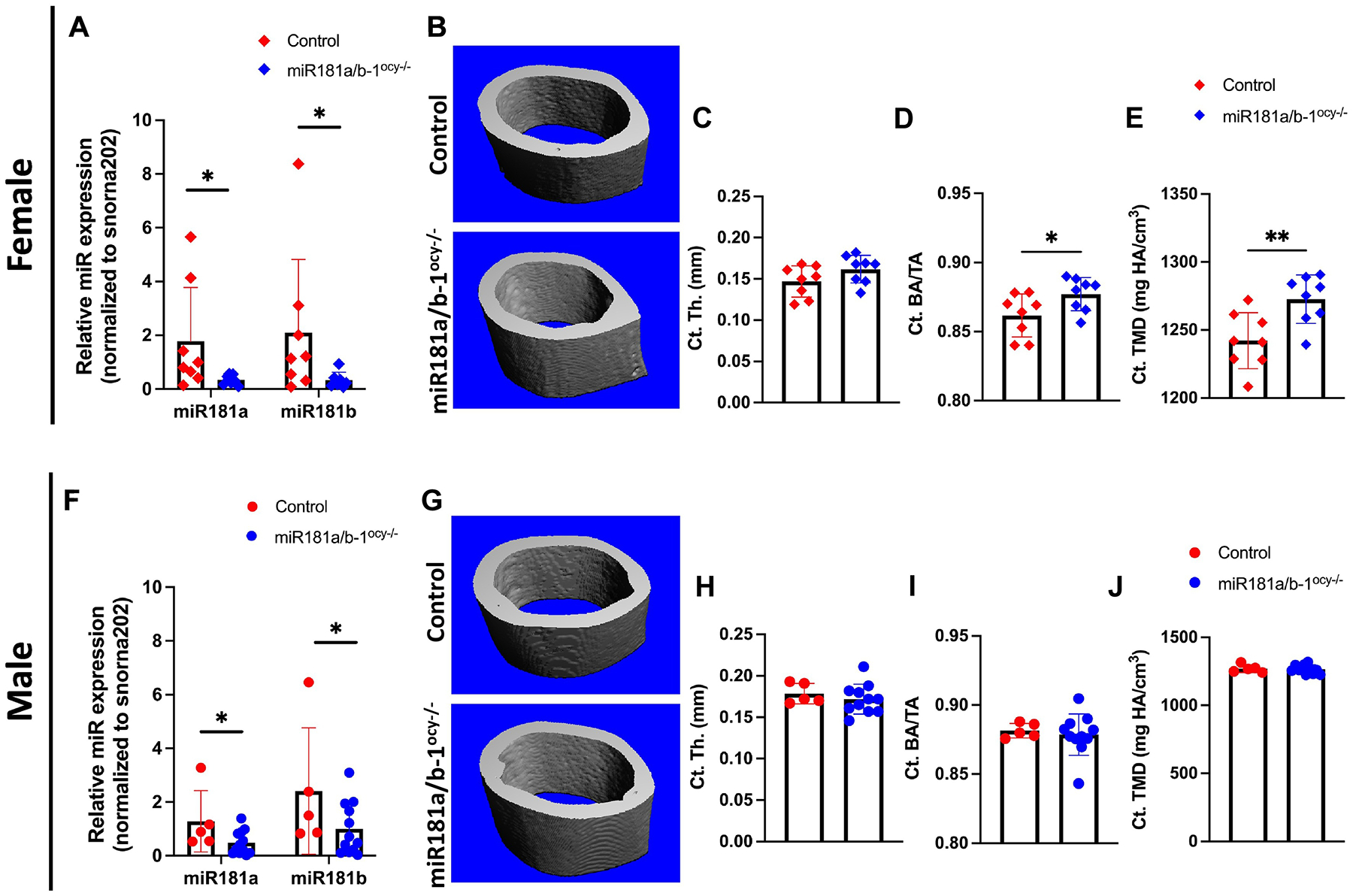
Bone derives its ability to resist fracture from bone mass and quality concurrently; however, many questions about the molecular mechanisms controlling bone quality remain unanswered, limiting the development of diagnostics and therapeutics. Despite the increasing evidence on the importance of miR181a/b-1 in bone homeostasis and disease, whether and how osteocyte-intrinsic miR181a/b-1 controls bone quality remains elusive. Osteocyte-intrinsic deletion of miR181a/b-1 in osteocytes in vivo resulted in compromised overall bone mechanical behavior in both sexes, although the parameters affected by miR181a/b-1 varied distinctly based on sex. Furthermore, impaired fracture resistance in both sexes was unexplained by cortical bone morphology, which was altered in female mice and intact in male mice with miR181a/b-1-deficient osteocytes. The role of miR181a/b-1 in the regulation of osteocyte metabolism was apparent in bioenergetic testing of miR181a/b-1-deficient OCY454 osteocyte-like cells and transcriptomic analysis of cortical bone from mice with osteocyte-intrinsic ablation of miR181a/b-1. Altogether, this study demonstrates the control of osteocyte bioenergetics and the sexually dimorphic regulation of cortical bone morphology and mechanical properties by miR181a/b-1, hinting at the role of osteocyte metabolism in the regulation of mechanical behavior.

Keywords: Bone quality; Cell metabolism; Mechanical behavior; Microrna; Osteocyte.


Conflict of interest statement

Declaration of competing interest The authors have no conflict of interest to declare.

Figures

Fig. 1.
Fig. 1.
miR181a/b-1 regulates metabolic genes and targets Sirt1. Compared to primary bone marrow stromal cells (pBMSCs, week 0), mature osteocytes differentiated from pBMSCs (week 3) exhibited increased miR181a/b-1 expression (A). Data represent n = 3 samples (mice used for primary cell isolation)/group. RT-qPCR revealed upregulated mRNA expression of Sirt1, Pten, and Cox11 with miR181a/b-1 inhibition in OCY454 osteocyte-like cells (B). Data represent 7 separate biological specimens collected across n = 3 independent experiments. miR181a/b-1 inhibition in OCY454 cells induced Sirt1 and Pten protein expression as measured by western blot (C-D). Data represent 11 separate biological specimens collected across n = 3 independent experiments. Sirt1 is a direct miR181a/b-1 target, as shown in a representative relative luciferase assay of OCY454 osteocyte-like cells transfected with Psicheck2-Sirt1 3′UTR and negative control or miR181a/b-1 inhibitors (E). Graph shows biological replicates of a representative experiment from n = 3 independent experiments, 3–5 biological replicates/experiment. *p < 0.05, **p < 0.01, ****p < 0.0001.
Fig. 2.
Fig. 2.
miR181a/b-1 modulates osteocyte bioenergetics in vitro. OCY454 osteocyte-like cells transfected with miR181a/b-1 inhibitors demonstrated reduced oxygen consumption rate (OCR, A-E), a measure of oxidative phosphorylation, compared to cells transfected with negative control inhibitors. Specifically, basal respiration (B) and ATP-linked respiration (C) were diminished due to miR181a/b-1 inhibition in osteocytes. Conversely, miR181a/b-1 deficiency did not alter any of the glycolysis parameters measured by extracellular acidification rate (ECAR, F–J). Data represent average of biological replicates from each independent experiment; n = 3 independent experiments, 4–5 biological replicates/experiment. *p < 0.05.
Fig. 3.
Fig. 3.
Osteocytic-intrinsic miR-181a/b-1 deficiency increases mineralization in female mouse bones. 8-week-old miR181a/b-1ocy−/− mice of both sexes exhibited reduced miR181a/b-1 expression in osteocyte-enriched cortical bone (A, F). n = 8 mice/group for females, n = 5–11 mice/group for males. microCT analysis of cortical bone from miR181a/b-1 ablated female mice (B) revealed increased cortical bone area fraction (Ct. BA/TA, D) and bone tissue mineral density (Ct. TMD, E) without significant changes in cortical thickness (Ct. Th, C). n = 8 mice/group for females. Cortical bone parameters remained unaffected by miR181a/b-1 ablation in males (G–J). n = 5–11 mice/group for males. *p < 0.05, **p < 0.01.
Fig. 4.
Fig. 4.
miR-181a/b-1 deletion in osteocytes compromises fracture resistance in both sexes. Load-displacement curves of female (A) and male (B) WT and miR181a/b-1ocy−/− femurs. Three-point bending testing performed on female 8-week-old miR181a/b-1ocy−/− mice demonstrated reduced work-to-fracture (D) and post-yield displacement (E) compared to their respective controls without changes in yield force (C), ultimate force (F), or elastic modulus (G). n = 8 mice/group for females. Ablation of osteocytic miR181a/b-1 in male mice suppressed yield force (H) while other parameters, such as work-to-fracture and post-yield displacement, remained unaffected (I–L). n = 5–11 mice/group for males. *p < 0.05, **p < 0.01.
Fig. 5.
Fig. 5.
Lacunocanalicular network remains unaffected by miR181a/b-1 inhibition. Box on representative 40× images indicating the approximate location of 100× Z-stack images acquired (A). Lacunocanalicular network is visualized with 3D reconstructed, maximum projections of cortical bone stained with DAPI to detect DNA, phalloidin to detect actin, and DiI to detect membranes (A). Representative merged RGB, maximum Z-projection images of whole mount cortical bone of female and male 8-week-old miR181a/b-1ocy−/− and control mice (B–E). Analysis revealed that canalicular tortuosity and osteocyte number remained intact in both females (F–G) and males (H–I). n = 5–6 mice/group for females and n = 4–6 mice/group for males.
Fig. 6.
Fig. 6.
miR181a/b-1 downregulates gene sets associated with mitochondrial function and metabolism in vivo. A heatmap of differentially expressed genes identified in cortical bone RNA from female (A) and male (B) miR181a/b-1ocy−/− mice and their respective controls. Greb1l mRNA expression remained unaffected in miR181a/b-1 inhibited OCY454 osteocyte-like cells compared to the controls (C) while miR181a/b-1 deficiency induced Lrmp mRNA expression (D) and a 4-fold upregulation of Myo15 mRNA expression (E). Data represent 12 separate biological specimens collected across n = 4 independent experiments. *p < 0.05, **p < 0.01. Gene-set enrichment analysis of transcriptomic analysis of cortical bone RNA from 8-week-old female and male miR181a/b-1ocy−/− mice and their respective controls representing up- and down-regulated gene sets (F).

References

    1. Wainwright SA, Marshall LM, Ensrud KE, Cauley JA, Black DM, Hillier TA, et al., Hip fracture in women without osteoporosis, J. Clin. Endocrinol. Metab 90 (5) (2005) 2787–2793. Epub 2005/02/25, 10.1210/jc.2004-1568. Epub 2005/02/25. - DOI - PubMed
    1. Schwartz AV, Vittinghoff E, Bauer DC, Hillier TA, Strotmeyer ES, Ensrud KE, et al., Association of BMD and FRAX score with risk of fracture in older adults with type 2 diabetes, JAMA 305 (21) (2011) 2184–2192. Epub 2011/06/03, 10.1001/jama.2011.715. Epub 2011/06/03. - DOI - PMC - PubMed
    1. Chang JL, Brauer DS, Johnson J, Chen CG, Akil O, Balooch G, et al., Tissue specific calibration of extracellular matrix material properties by transforming growth factor-beta and Runx2 in bone is required for hearing, EMBO Rep. 11 (10) (2010) 765–771. Epub 2010/09/18, 10.1038/embor.2010.135. Epub 2010/09/18. - DOI - PMC - PubMed
    1. Kawashima Y, Fritton JC, Yakar S, Epstein S, Schaffler MB, Jepsen KJ, et al., Type 2 diabetic mice demonstrate slender long bones with increased fragility secondary to increased osteoclastogenesis, Bone 44 (4) (2009) 648–655. Epub 2009/01/20, 10.1016/j.bone.2008.12.012. Epub 2009/01/20. - DOI - PMC - PubMed
    1. Tommasini SM, Nasser P, Jepsen KJ, Sexual dimorphism affects tibia size and shape but not tissue-level mechanical properties, Bone. 40 (2) (2007) 498–505. Epub 2006/10/13, 10.1016/j.bone.2006.08.012. Epub 2006/10/13. - DOI - PubMed

Publication types