. 2023 Sep;215(3):107995.
doi: 10.1016/j.jsb.2023.107995. Epub 2023 Jul 4.

Double-headed binding of myosin II to F-actin shows the effect of strain on head structure

Affiliations

Double-headed binding of myosin II to F-actin shows the effect of strain on head structure

Alimohammad Hojjatian et al. J Struct Biol. 2023 Sep.

Abstract

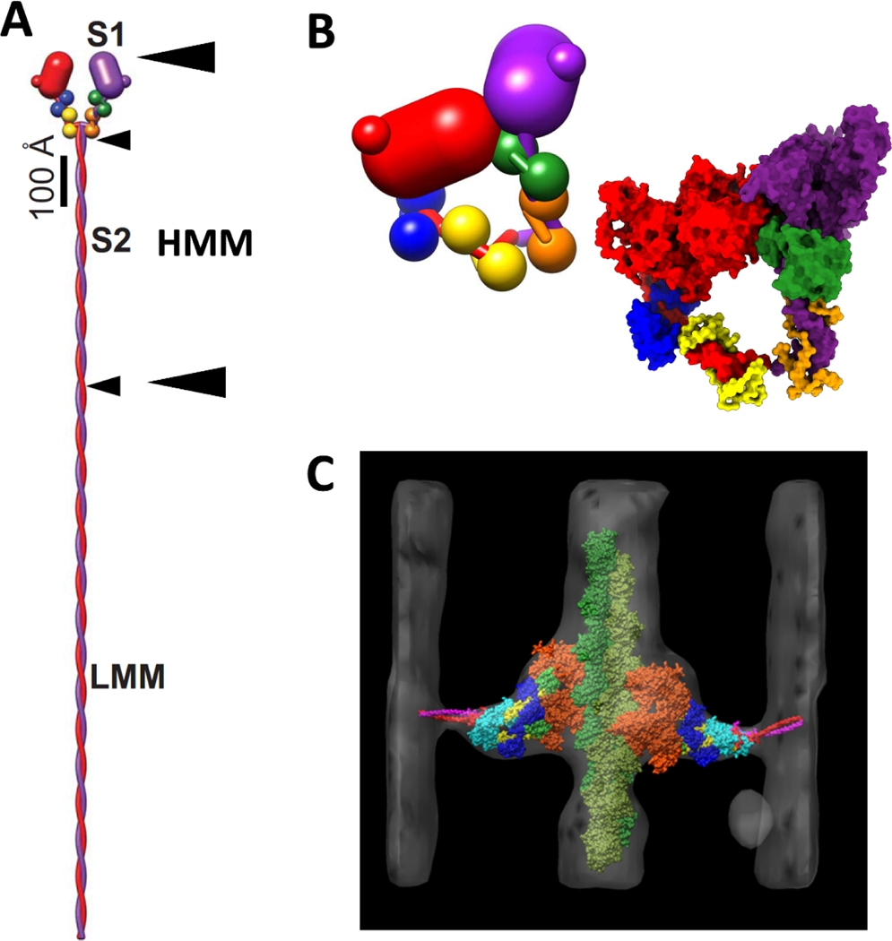
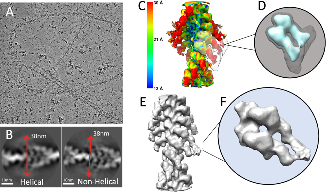
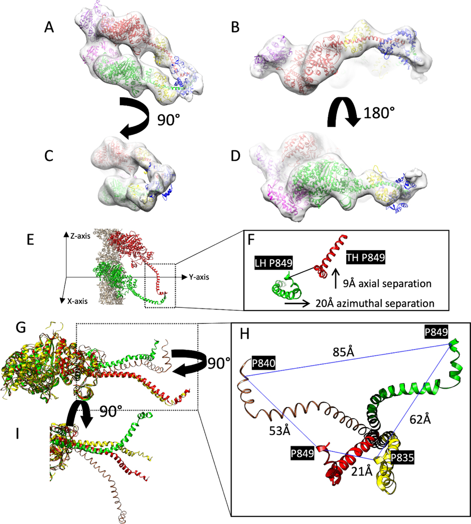
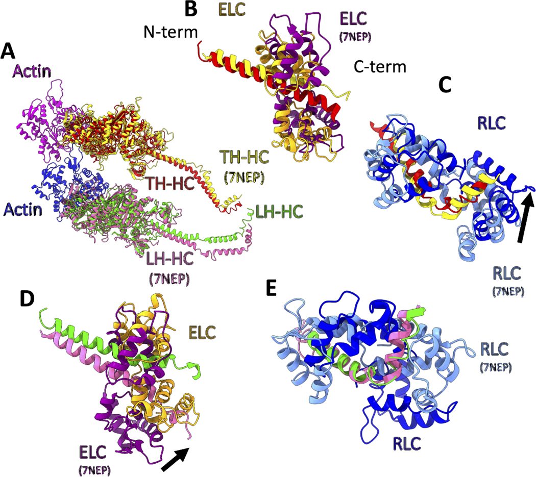
Force production in muscle is achieved through the interaction of myosin and actin. Strong binding states in active muscle are associated with Mg·ADP bound to the active site; release of Mg·ADP allows rebinding of ATP and dissociation from actin. Thus, Mg·ADP binding is positioned for adaptation as a force sensor. Mechanical loads on the lever arm can affect the ability of myosin to release Mg·ADP but exactly how this is done is poorly defined. Here we use F-actin decorated with double-headed smooth muscle myosin fragments in the presence of Mg·ADP to visualize the effect of internally supplied tension on the paired lever arms using cryoEM. The interaction of the paired heads with two adjacent actin subunits is predicted to place one lever arm under positive and the other under negative strain. The converter domain is believed to be the most flexible domain within myosin head. Our results, instead, point to the segment of heavy chain between the essential and regulatory light chains as the location of the largest structural change. Moreover, our results suggest no large changes in the myosin coiled coil tail as the locus of strain relief when both heads bind F-actin. The method would be adaptable to double-headed members of the myosin family. We anticipate that the study of actin-myosin interaction using double-headed fragments enables visualization of domains that are typically noisy in decoration with single-headed fragments.

Keywords: Filaments; Helical reconstruction; Lipid monolayer; Single particle reconstruction; Structural adjustments to strain; Structure heterogeneity.


Conflict of interest statement

Declaration of Competing Interest The authors declare that they have no known competing financial interests or personal relationships that could have appeared to influence the work reported in this paper.

Figures

Figure 1.
Figure 1.
Myosin II structure and conformations. (A) Myosin hexamer with two heavy chains, red and purple, and two pairs of light chains, ELC, blue and green, and RLC yellow and orange. Arrow heads mark the boundaries for the S1, S2, HMM and LMM subfragments. The small arrow heads mark the length of S2 and LMM. The pair of large arrowheads mark the boundaries of HMM. (B) The interacting heads motif in which one head, dubbed the blocked head, red, binds the second head, dubbed the free head, purple. Insert at the lower right is a space-filling rendition using PDB 1i84. (C) Atomic model of a 2-headed, rigor myosin cross-bridge from electron tomograms of swollen Lethocerus flight muscle from Liu et al. (2006) showing convergence toward the head-tail junction. In this orientation, the Z-line would be at the bottom and the M-line at the top. (A,B) Adapted from Hu et al. (2016).
Figure 2.
Figure 2.
Cryo-EM image processing of F-actin decorated with smooth muscle HMM. (A) An electron micrograph showing F-actin decorated with HMM. (B) Showing representative good class averages resulted from 2D classification in Relion performed with and without imposed helical symmetry. Red arrows show an estimate of the diameter of the filaments to be approximately 380Å. (C) Result of local resolution determination using Local MonoRes (Vilas et al., 2018) shown as a heatmap of the refinement after selecting the best classes from unmasked 3D classifications. (D) The 3D soft-edge mask (shown in grey) used for 3D classifications. This mask includes both motor domains and the blurred lever arms (shown in cyan). (E) The best Acto-HMM density map, 3D reconstructed by only including the segments from group 1. (F) Magnified view of the segmented map in E.
Figure 3.
Figure 3.
Comparison of atomic models of HMM decorated F-actin with closely similar structures. These are rigid body fits of the S1 atomic structure into the reconstruction. (A-D) Different views of the acto-HMM atomic model in the density map of Leading and Trailing heads. The two adjacent actin subunits are shown in purple and magenta, heavy chains of the Leading and Trailing heads are shown in green and red; the ELCs and RLCs are shown in blue and yellow, respectively. (E,F) Showing the distance between the conserved pro849 residues from the Leading Head, LH, and the Trailing Head TH, Z-axis showing the direction along the axis of the filament. (G,H) different views of the comparison of the heavy chains from the Trailing Head (red) and Leading Head (green), the crystal structure of scallop myosin S1 in the near rigor conformation (PDB ID: 1KK7) (yellow) and the cryo-EM structure of chicken skeletal S1 in the rigor state (brown) after the alignment of the motor domains. The distances are measured between the invariant proline residues. (I) Axial view showing the conformational difference between Trailing Head (red), Leading Head (green) and the starting model 1KK7 (yellow) after the alignment of the ELC binding segment of the lever arm.
Figure 4.
Figure 4.
Comparison of the smooth muscle acto-HMM atomic model with that for skeletal muscle myosin II (PDB ID: 7NEP) obtained by cryoET (Wang et al., 2021). The heavy chains from the two models have been aligned at the motor domains. (A) shows the alignment of the heavy chains from the two models and the actin subunits (magenta and medium blue). Heavy chains of Trailing head (red and yellow for smooth and skeletal muscle myosin, respectively) and Leading heads (green and hot pink for smooth and skeletal muscle myosin, respectively). (B) Comparison of ELCs and the ELC binding domains of the Trailing Head. Skeletal ELC and heavy chain (purple and yellow respectively) and smooth ELC and heavy chain (orange and red respectively). (C) Comparison of the RLCs and the RLC binding domains of the Trailing Head. Skeletal and smooth RLCs are shown in cornflower blue and blue, respectively. The black arrow shows the direction of the movement needed for alignment of the skeletal RLC to the smooth RLC model. (D) Similar to (B), showing ELCs for the Leading Head. The difference in the conformation of the two heads is more significant in the Leading Head. (E) Similar to (C), showing RLCs for the Leading Head, after the alignment of the heavy chain residues in the RLC binding domains.

Similar articles

References

    1. Afonine PV, Klaholz BP, Moriarty NW, Poon BK, Sobolev OV, Terwilliger TC, Adams PD, Urzhumtsev A, 2018. New tools for the analysis and validation of cryo-EM maps and atomic models. Acta Crystallogr D Struct Biol 74, 814–840. - PMC - PubMed
    1. Arakelian C, Warrington A, Winkler H, Perz-Edwards RJ, Reedy MK, Taylor KA, 2015. Myosin S2 origins track evolution of strong binding on actin by azimuthal rolling of motor domain. Biophys J 108, 1495–1502. - PMC - PubMed
    1. Banerjee C, Hu Z, Huang Z, Warrington JA, Taylor DW, Trybus KM, Lowey S, Taylor KA, 2017. The structure of the actin-smooth muscle myosin motor domain complex in the rigor state. J Struct Biol 200, 325–333. - PMC - PubMed
    1. Behrmann E, Muller M, Penczek PA, Mannherz HG, Manstein DJ, Raunser S, 2012. Structure of the rigor actin-tropomyosin-myosin complex. Cell 150, 327–338. - PMC - PubMed
    1. Blankenfeldt W, Thoma NH, Wray JS, Gautel M, Schlichting I, 2006. Crystal structures of human cardiac beta-myosin II S2-Delta provide insight into the functional role of the S2 subfragment. Proc Natl Acad Sci U S A 103, 17713–17717. - PMC - PubMed

Publication types

MeSH terms

LinkOut - more resources