. 2023 Jul 6;13(1):10969.
doi: 10.1038/s41598-023-37966-5.

Extracellular vesicle analysis of plasma allows differential diagnosis of atypical pancreatic serous cystadenoma

Affiliations

Extracellular vesicle analysis of plasma allows differential diagnosis of atypical pancreatic serous cystadenoma

Katherine S Yang et al. Sci Rep. .

Abstract

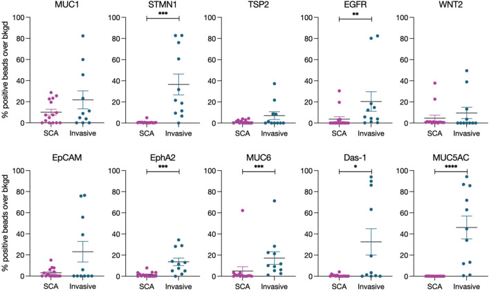
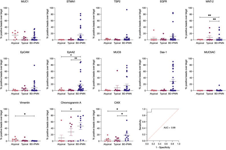
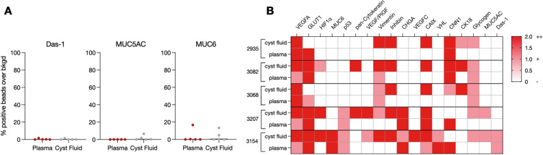
Increased use of cross-sectional imaging has resulted in frequent detection of incidental cystic pancreatic lesions. Serous cystadenomas (SCAs) are benign cysts that do not require surgical intervention unless symptomatic. Unfortunately, up to half of SCAs do not have typical imaging findings ("atypical SCAs"), overlap with potentially malignant precursor lesions, and thus pose a diagnostic challenge. We tested whether the analysis of circulating extracellular vesicle (EV) biomarkers using a digital EV screening technology (DEST) could enhance the discrimination of cystic pancreatic lesions and avoid unnecessary surgical intervention in these atypical SCAs. Analysis of 25 different protein biomarkers in plasma EV from 68 patients identified a putative biomarker signature of Das-1, Vimentin, Chromogranin A, and CAIX with high discriminatory power (AUC of 0.99). Analysis of plasma EV for multiplexed markers may thus be helpful in clinical decision-making.


Conflict of interest statement

RW is a consultant to ModeRNA, Lumicell, Seer Biosciences, Earli, and Accure Health, none of whom contributed to this research. The other authors have no competing interest.

Figures

Figure 1
Figure 1
Differential diagnosis of atypical SCA. Atypical SCA can be mistaken for other pancreatic cysts (MCN, BD-IPMN), some of which have malignant potential (left panel). Plasma EV analysis using a digital EV screening technology (DEST, right) represents a new approach for the identification and management of atypical SCA. The DEST assay measures EV biomarkers using a magnetic bead amplification technique. EV containing a biomarker of interest are captured on an antibody-coated bead, thus concentrating EV magnetically. Biotinylated detection antibodies and subsequent tyramide signal amplification is used to read out positive beads by imaging (illustrative example bottom right) or flow cytometry (readout for this manuscript). An example of positive DEST beads is shown in the lower right, with single beads colored purple and positive beads from tyramide signal amplification colored cyan. Images were generated using ImageJ2, version 2.9.0/1.53t (https://imagej.net/software/fiji/).
Figure 2
Figure 2
EV analysis allows the detection of invasive cancers in cystic lesions. SCA (purple; all cases of typical and atypical SCA by imaging) plasma EV were compared to invasive IPMN (blue) using the indicated biomarkers. Error bars represent standard error of the mean. P-values < 0.05 (*) were calculated using a Mann–Whitney test.
Figure 3
Figure 3
Biomarker expression in atypical and typical SCA compared to BD-IPMN. Atypical (light pink) and typical (dark pink) SCA compared to BD-IPMN (dark purple) for malignant and benign biomarkers. Error bars represent standard error of the mean. P-values (*, < 0.05) were determined using a Mann–Whitney test. Multiple logistic regression analysis of atypical versus BD-IPMN using a four-way biomarker combination of Das-1, Vimentin, Chromogranin A, and Carbonic Anhydrase I × (CAIX) is shown in the bottom right ROC curve. The AUC was determined using Prism.
Figure 4
Figure 4
SCA DEST plasma correlation with cyst fluid. Biomarkers were measured in matched SCA plasma and cyst fluid samples from five patients. (A) Malignant biomarkers are at low expression in both plasma and cyst fluid. (B) SCA biomarker correlation between matched plasma and cyst fluid samples.

References

    1. Zerboni G, et al. Systematic review and meta-analysis: Prevalence of incidentally detected pancreatic cystic lesions in asymptomatic individuals. Pancreatology. 2019;19:2–9. doi: 10.1016/j.pan.2018.11.014. - DOI - PubMed
    1. Brugge WR, Lauwers GY, Sahani D, Fernandez-del CC, Warshaw AL. Cystic neoplasms of the pancreas. N. Engl. J. Med. 2004;351:1218–1226. doi: 10.1056/NEJMra031623. - DOI - PubMed
    1. Roldán J, et al. Evolving trends in pancreatic cystic tumors: A 3-decade single-center experience with 1290 resections. Ann. Surg. 2023;277:491–497. doi: 10.1097/SLA.0000000000005142. - DOI - PubMed
    1. Van Dyke TJ, Johlin FC, Bellizzi AM, Howe JR. Serous cystadenocarcinoma of the pancreas: Clinical features and management of a rare tumor. Dig. Surg. 2016;33:240–248. doi: 10.1159/000444721. - DOI - PubMed
    1. Lombardo C, et al. Incidence and reasons of pancreatic resection in patients with asymptomatic serous cystadenoma. Pancreatology. 2018;18:577–584. doi: 10.1016/j.pan.2018.06.001. - DOI - PubMed

Publication types