. 2023 Jul 6;13(1):10960.
doi: 10.1038/s41598-023-38152-3.

ASC- and caspase-1-deficient C57BL/6 mice do not develop demyelinating disease after infection with Theiler's murine encephalomyelitis virus

Affiliations

ASC- and caspase-1-deficient C57BL/6 mice do not develop demyelinating disease after infection with Theiler's murine encephalomyelitis virus

Dandan Li et al. Sci Rep. .

Abstract

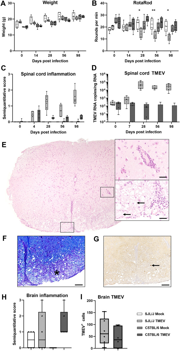
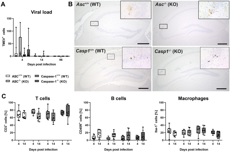
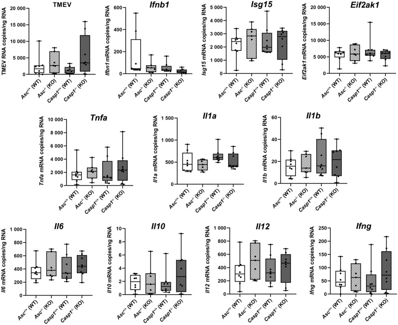
Theiler's murine encephalomyelitis virus (TMEV) induces an acute polioencephalomyelitis and a chronic demyelinating leukomyelitis in SJL mice. C57BL/6 (B6) mice generally do not develop TMEV-induced demyelinating disease (TMEV-IDD) due to virus elimination. However, TMEV can persist in specific immunodeficient B6 mice such as IFNβ-/- mice and induce a demyelinating process. The proinflammatory cytokines IL-1β and IL-18 are activated by the inflammasome pathway, which consists of a pattern recognition receptor molecule sensing microbial pathogens, the adaptor molecule Apoptosis-associated speck-like protein containing a CARD (ASC), and the executioner caspase-1. To analyze the contribution of the inflammasome pathway to the resistance of B6 mice to TMEV-IDD, ASC- and caspase-1-deficient mice and wild type littermates were infected with TMEV and investigated using histology, immunohistochemistry, RT-qPCR, and Western Blot. Despite the antiviral activity of the inflammasome pathway, ASC- and caspase-1-deficient mice eliminated the virus and did not develop TMEV-IDD. Moreover, a similar IFNβ and cytokine gene expression was found in the brain of immunodeficient mice and their wild type littermates. Most importantly, Western Blot showed cleavage of IL-1β and IL-18 in all investigated mice. Consequently, inflammasome-dependent activation of IL-1β and IL-18 does not play a major role in the resistance of B6 mice to TMEV-IDD.


Conflict of interest statement

The authors declare no competing interests.

Figures

Figure 1
Figure 1
Clinical and histological data of Asc−/− and Casp1−/− mice (KO) and wild type littermates (WT) infected with 1 × 105 PFU of the BeAn strain of Theiler’s murine encephalomyelitis virus. Weight analysis (A) and Rotarod performance tests (B) showed a continuous increase in body weight and no deterioration in motor coordination in all mice. Semiquantitative scores of perivascular mononuclear cell infiltrates in the brain (C) and spinal cord (D) at 4, 14 and 98 days post infection (dpi). Cell infiltrates were analyzed in two complete cross sections of the brain at the levels of the hippocampus and cerebellum and complete cross sections of the cervical, thoracic and lumbar spinal cord. Mild to moderate inflammation was found at 4 and 14 dpi but not at 98 dpi in the brain, whereas mild inflammation was present in the spinal cord of at all investigated time points. No significant differences between KO and WT mice in the clinical and histological data were detected using Mann–Whitney tests. n = 7–10. Box plots with all data points. (E) Images demonstrate few perivascular mononuclear cells in the hippocampus and spinal cord of all mice at 4 dpi. Hematoxylin and eosin (HE) staining. Bars = 400 µm.
Figure 2
Figure 2
Clinical disease and histological lesions in SJL/J and C57BL/6 mice mock-infected with cell culture medium or infected with 1.63 × 106 PFU of the BeAn strain of Theiler’s murine encephalomyelitis virus (TMEV). Weight analysis (A) and Rotarod performance tests (B) of TMEV-infected SJL/J showed a continuous increase in body weight but an impaired motor coordination at 28, 56, and 98 days post infection (dpi). No clinical signs were present in C57BL/6 mice. Mann–Whitney tests: *P < 0.05; **P < 0.01. n = 6. (C) Semiquantitative scores of perivascular mononuclear cell infiltrates analyzed in complete cross sections of the cervical, thoracic and lumbar spinal cord. Severe inflammation only develops in SJL/J mice. n = 5–6. (D) TMEV RNA copy numbers in the spinal cord quantified with RT-qPCR. Note the logarithmic scale and high versus low/no virus replication in SJL/J and C57BL/6 mice at 28, 56 and 98 dpi, respectively. n = 5–6. (E) Mononuclear cell infiltrates in the cervical spinal cord of a TMEV-infected SJL/J mouse at 98 dpi. Note perivascular mononuclear cells and gitter cells (arrows) in higher magnifications. Hematoxylin and eosin (HE) staining. Bars = 30 µm. (F) Demyelination in the inflamed ventrolateral funiculus of the spinal cord (same SJL/J mouse as in (F)). Note loss of luxol fast blue (LFB) staining due to severe inflammation (asterisk). Bar = 100 µm. (G) Immunohistochemistry was used to detect virus antigen in the spinal cord (same mouse as in (E) and (F)). Note TMEV+ cell (arrow) in the spinal cord white matter lesion. Avidin–biotin-complex (ABC) method using 3,3'-diaminobenzidine (DAB) as chromogen. Bar = 100 µm. (H) Semiquantitative scores of perivascular mononuclear cell infiltrates in the brain analyzed in a complete cross section of the cerebrum at the level of the hippocampus at 4 dpi. Moderate to strong inflammatory lesions were found in TMEV-infected SJL/J and C57BL/6 mice. (I) Immunohistochemistry was used to detect virus antigen in a complete cross section of the cerebrum at the level of the hippocampus at 4 dpi. No significant differences in the number of TMEV+ cells were found between SJL/J and C57BL/6 mice at 4 dpi using Mann–Whitney tests. n = 6. Box plots with all data points.
Figure 3
Figure 3
Immunohistochemistry was used to detect virus antigen and determine percentages of perivascular immune cells in the brain of Asc−/− and Casp1−/− mice (KO) and wild type littermates (WT) infected with 1 × 105 PFU of the BeAn strain of Theiler’s murine encephalomyelitis virus (TMEV). (A) All TMEV+ cells were counted in a complete cross section of the cerebrum at the level of the hippocampus at 4, 14 and 98 days post infection (dpi). No significant differences in the number of TMEV+ cells were found between KO and WT mice using Mann–Whitney tests demonstrating rapid virus elimination. n = 7–10. (B) Images show TMEV+ cells in the CA1 and CA2 area of the hippocampus of KO and WT mice at 4 dpi. Avidin–biotin-complex (ABC) method using 3,3'-diaminobenzidine (DAB) as chromogen. Bars = 400 µm. (C) Percentages of perivascular CD3+ T cell, CD45R+ B cells and Iba-1+ macrophages at 4 and 7 dpi. No significant differences between KO and WT mice were detected using Mann–Whitney tests. n = 10 except Casp1−/− mice at 4 dpi (n = 9). Box plots with all data points.
Figure 4
Figure 4
The number of Theiler’s murine encephalomyelitis virus (TMEV) RNA copies/ng RNA as well as Ifnb1, Isg15, Eif2ak1 (PKR), Tnfa, Il1a, Il1b, Il6, Il10, Il12 (p40) and Ifng mRNA copies /ng RNA was quantified in the brain of Asc−/− and Casp1−/− mice (KO) and wild type littermates (WT) infected with 1 × 105 PFU of the BeAn strain of TMEV at 4 days post infection using RT-qPCR. No significant differences between Asc−/− and Casp1−/− mice and their respective wild type littermates were found using Mann–Whitney tests. n = 8 (Asc+/+, Casp1+/+ and Casp1−/− mice), n = 7 (Asc−/− mice). Box plots with all data points.
Figure 5
Figure 5
Western Blot was used to detect cleavage of the inactive precursors of the proinflammatory cytokines in the brain of Asc−/− and Casp1−/− mice (KO) and wild type littermates (WT) infected with 1 × 105 PFU of the BeAn strain of Theiler’s murine encephalomyelitis virus (TMEV). Brain samples of SJL/J and C57BL/6 mice mock-infected with cell culture medium or infected with 1.63 × 106 PFU of the TMEV-BeAn were also included. (A) A cleavage of the precursor Pro-IL-1β to the mature IL-1β protein was found in all brain samples. (B) Similarly, a cleavage of the precursor Pro-IL-18 to the mature IL-18 protein was demonstrated in all brain samples despite low protein levels in Asc−/− mice. Detection of Lamin B1 served as the loading control.

Similar articles

Cited by

References

    1. Theiler M. Spontaneous encephalomyelitis of mice, a new virus disease. J. Exp. Med. 1937;65:705–719. doi: 10.1084/jem.65.5.705. - DOI - PMC - PubMed
    1. Mecha M, Carrillo-Salinas FJ, Mestre L, Feliu A, Guaza C. Viral models of multiple sclerosis: Neurodegeneration and demyelination in mice infected with Theiler's virus. Prog. Neurobiol. 2013;101–102:46–64. doi: 10.1016/j.pneurobio.2012.11.003. - DOI - PMC - PubMed
    1. Oleszak EL, Chang JR, Friedman H, Katsetos CD, Platsoucas CD. Theiler's virus infection: A model for multiple sclerosis. Clin. Microbiol. Rev. 2004;17:174–207. doi: 10.1128/CMR.17.1.174-207.2004. - DOI - PMC - PubMed
    1. Tsunoda I, et al. Three immune-mediated disease models induced by Theiler's virus: Multiple sclerosis, seizures and myocarditis. Clin. Exp. Neuroimmunol. 2016;7:330–345. doi: 10.1111/cen3.12341. - DOI - PMC - PubMed
    1. Tsunoda I, Fujinami RS. Neuropathogenesis of Theiler's murine encephalomyelitis virus infection, an animal model for multiple sclerosis. J. Neuroimmune Pharmacol. 2010;5:355–369. doi: 10.1007/s11481-009-9179-x. - DOI - PMC - PubMed

Publication types

LinkOut - more resources