. 2023 Dec;28(12):5350-5358.
doi: 10.1038/s41380-023-02157-1. Epub 2023 Jul 6.

Age-related vulnerability of the human brain connectome

Affiliations

Age-related vulnerability of the human brain connectome

Massimo Filippi et al. Mol Psychiatry. 2023 Dec.

Abstract

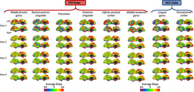
Multifactorial models integrating brain variables at multiple scales are warranted to investigate aging and its relationship with neurodegeneration. Our aim was to evaluate how aging affects functional connectivity of pivotal regions of the human brain connectome (i.e., hubs), which represent potential vulnerability 'stations' to aging, and whether such effects influence the functional and structural changes of the whole brain. We combined the information of the functional connectome vulnerability, studied through an innovative graph-analysis approach (stepwise functional connectivity), with brain cortical thinning in aging. Using data from 128 cognitively normal participants (aged 20-85 years), we firstly investigated the topological functional network organization in the optimal healthy condition (i.e., young adults) and observed that fronto-temporo-parietal hubs showed a highly direct functional connectivity with themselves and among each other, while occipital hubs showed a direct functional connectivity within occipital regions and sensorimotor areas. Subsequently, we modeled cortical thickness changes over lifespan, revealing that fronto-temporo-parietal hubs were among the brain regions that changed the most, whereas occipital hubs showed a quite spared cortical thickness across ages. Finally, we found that cortical regions highly functionally linked to the fronto-temporo-parietal hubs in healthy adults were characterized by the greatest cortical thinning along the lifespan, demonstrating that the topology and geometry of hub functional connectome govern the region-specific structural alterations of the brain regions.


Conflict of interest statement

The authors declare no conflict of interest in relation to the submitted work. Potential conflicts of interest outside the reported study are: MF is Editor-in-Chief of the Journal of Neurology, Associate Editor of Human Brain Mapping, Neurological Sciences, and Radiology; received compensation for consulting services from Alexion, Almirall, Biogen, Merck, Novartis, Roche, Sanofi; speaking activities from Bayer, Biogen, Celgene, Chiesi Italia SpA, Eli Lilly, Genzyme, Janssen, Merck-Serono, Neopharmed Gentili, Novartis, Novo Nordisk, Roche, Sanofi, Takeda, and TEVA; participation in Advisory Boards for Alexion, Biogen, Bristol-Myers Squibb, Merck, Novartis, Roche, Sanofi, Sanofi-Aventis, Sanofi-Genzyme, Takeda; scientific direction of educational events for Biogen, Merck, Roche, Celgene, Bristol-Myers Squibb, Lilly, Novartis, Sanofi-Genzyme; he receives research support from Biogen Idec, Merck-Serono, Novartis, Roche, Italian Ministry of Health, and Fondazione Italiana Sclerosi Multipla. CC, SB, EGS, VC, ML, and EC have nothing to disclose. FA is Associate Editor of NeuroImage: Clinical, has received speaker honoraria from Biogen Idec, Roche, Zambon and italfarmaco, and receives or has received research supports from the Italian Ministry of Health, Italian Ministry of University and Research, AriSLA (Fondazione Italiana di Ricerca per la SLA), the European Research Council and Foundation Research on Alzheimer Disease.

Figures

Fig. 1
Fig. 1. Stepwise functional connectivity average maps in young healthy adults.
Cortical maps represent characterization of stepwise connectivity analysis from the FTP and the OCC hubs in healthy young adults. Results are depicted in surface space per each of the eight hubs. Yellow/red areas represent strong functional connectivity with the hub, whereas blue/violet areas represent weak functional connectivity. FTP fronto-temporo-parietal, OCC occipital.
Fig. 2
Fig. 2. Differences between young and old healthy adults in stepwise functional connectivity of the eight hubs.
AH Cortical maps represent the significant differences in stepwise functional connectivity values between young healthy adults and older healthy adults. Statistical analysis was adjusted for sex and education. Results were corrected for multiple comparisons using a threshold-free cluster enhancement method combined with nonparametric permutation testing at p < 0.05 FWE-corrected. Color bars show the t-statistic applicable to the image. Red-yellow areas represent decreased functional connectivity in older adults relative to young adults, whereas blue/green areas represent enhanced functional connectivity in older adults compared to young.
Fig. 3
Fig. 3. Correlation analysis between combined SFC maps of young group and normalized cortical thickness values in older adults.
Relationship between combined SFC maps of the middle frontal gyrus (FTP hub) (A) and OCC hubs (B, C) and the cortical thickness of older adults normalized relative to young adults. FTP fronto-temporo-parietal, OCC occipital, SFC stepwise functional connectivity.
Fig. 4
Fig. 4. Boxplot of normalized cortical thickness values of highly connected regions with hubs in older healthy adults.
Cortical thickness value distribution of brain regions highly functionally connected to FTP hubs and OCC hubs. The red dotted line qualitatively highlights that cortical thickness values of the FTP-linked regions are lower than those of OCC-linked regions. FTP fronto-temporo-parietal, OCC occipital.

References

    1. Lopez-Otin C, Blasco MA, Partridge L, Serrano M, Kroemer G. The hallmarks of aging. Cell. 2013;153:1194–217. - PMC - PubMed
    1. Damoiseaux JS. Effects of aging on functional and structural brain connectivity. NeuroImage. 2017;160:32–40. - PubMed
    1. Terribilli D, Schaufelberger MS, Duran FL, Zanetti MV, Curiati PK, Menezes PR, et al. Age-related gray matter volume changes in the brain during non-elderly adulthood. Neurobiol Aging. 2011;32:354–68. - PMC - PubMed
    1. Salat DH, Buckner RL, Snyder AZ, Greve DN, Desikan RS, Busa E, et al. Thinning of the cerebral cortex in aging. Cereb Cortex. 2004;14:72–730. - PubMed
    1. Fjell AM, Walhovd KB. Structural brain changes in aging: courses, causes and cognitive consequences. Rev Neurosci. 2010;21:187–221. - PubMed

Publication types