. 2023 Jul 6;42(1):160.
doi: 10.1186/s13046-023-02714-0.

Cancer-associated fibroblast-derived PAI-1 promotes lymphatic metastasis via the induction of EndoMT in lymphatic endothelial cells

Affiliations

Cancer-associated fibroblast-derived PAI-1 promotes lymphatic metastasis via the induction of EndoMT in lymphatic endothelial cells

Wen-Fei Wei et al. J Exp Clin Cancer Res. .

Abstract

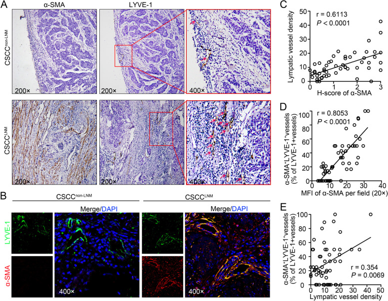
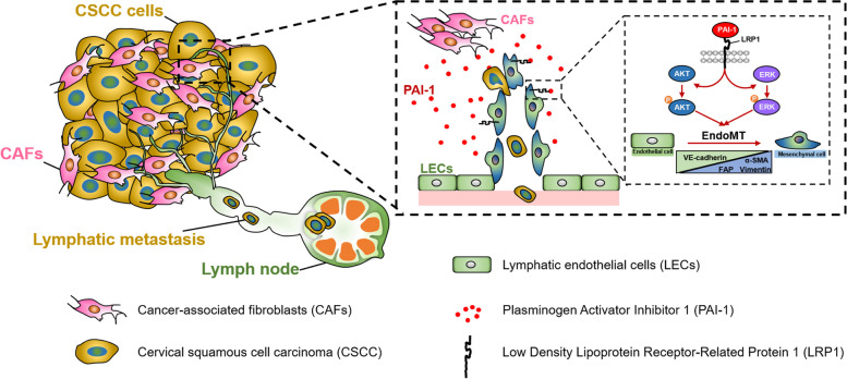
Background: Endothelial-mesenchymal transition (EndoMT) is an emerging adaptive process that modulates lymphatic endothelial function to drive aberrant lymphatic vascularization in the tumour microenvironment (TME); however, the molecular determinants that govern the functional role of EndoMT remain unclear. Here, we show that cancer-associated fibroblast (CAF)-derived PAI-1 promoted the EndoMT of lymphatic endothelial cells (LECs) in cervical squamous cell carcinoma (CSCC).

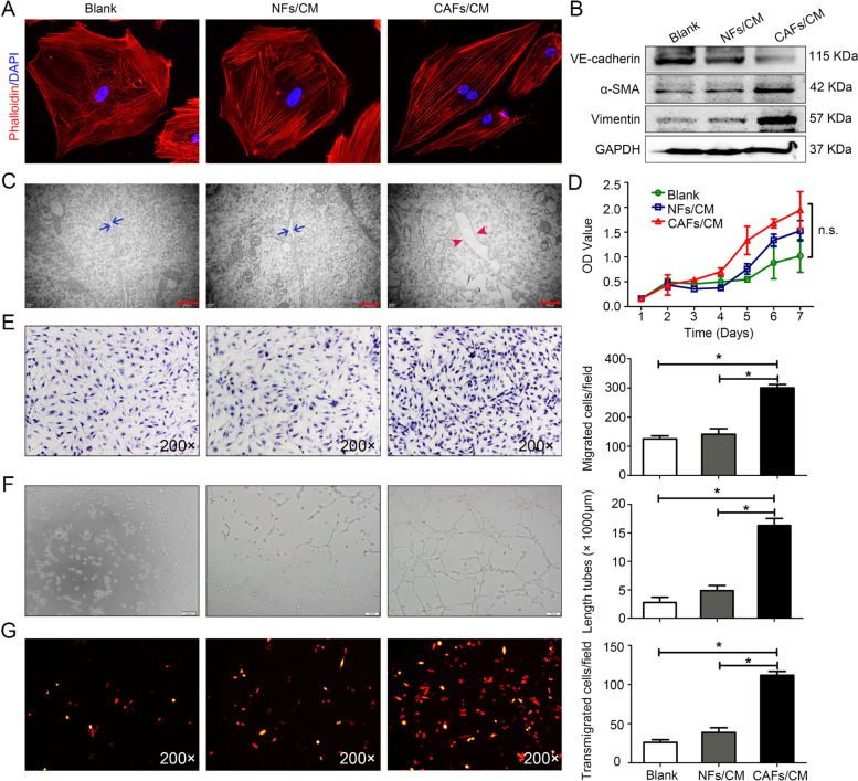
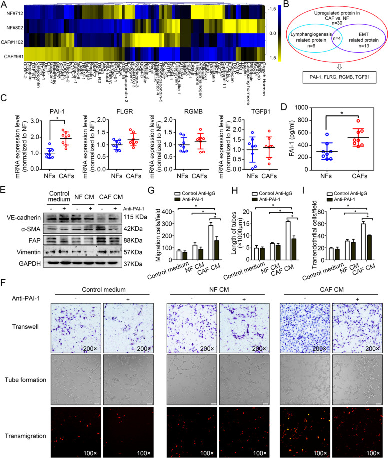
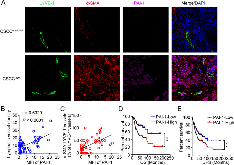
Methods: Immunofluorescent staining of α-SMA, LYVE-1 and DAPI were examined in primary tumour samples obtained from 57 CSCC patients. Assessment of cytokines secreted by CAFs and normal fibroblasts (NFs) was performed using human cytokine antibody arrays. The phenotype of EndoMT in lymphatic endothelial cells (LECs), gene expression levels, protein secretion and activity of signaling pathways were measured by real-time RT-PCR, ELISA or western blotting. The function of lymphatic endothelial monolayers was examined by transwell, tube formation assay, transendothelial migration assay in vitro. Lymphatic metastasis was measured using popliteal lymph node metastasis model. Furthermore, association between PAI-1 expression and EndoMT in CSCC was analyzed by immunohistochemistry. The Cancer Genome Atlas (TCGA) databases was used to assess the association of PAI-1 with survival rate in CSCC.

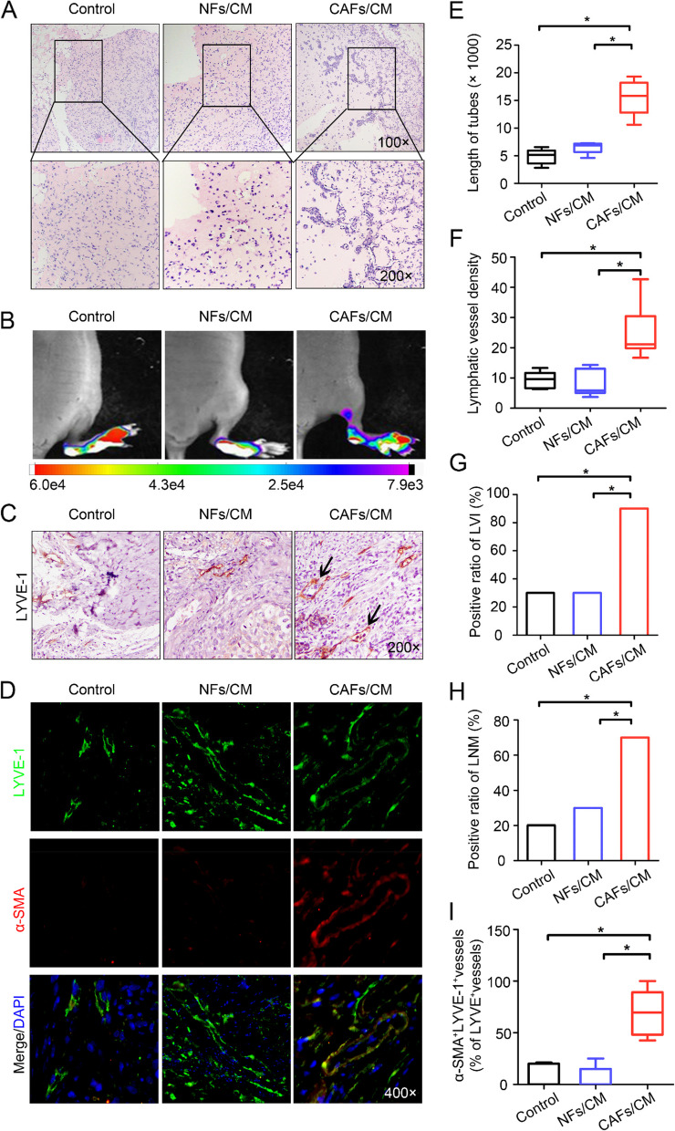
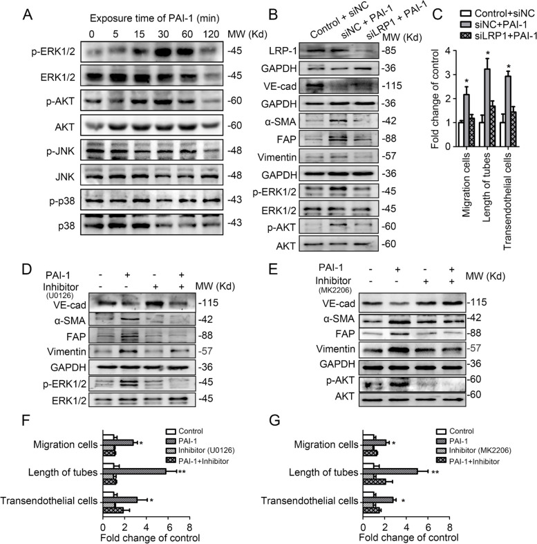
Results: CAF-derived PAI-1 promoted the EndoMT of LECs in CSCC. LECs undergoing EndoMT could initiate tumour neolymphangiogenesis that facilitated cancer cell intravasation/extravasation, which in turn promoted lymphatic metastasis in CSCC. Mechanistically, PAI-1 activated the AKT/ERK1/2 pathways by directly interacting with low-density lipoprotein receptor-related protein (LRP1), thereby leading to elevated EndoMT activity in LECs. Blockade of PAI-1 or inhibition of LRP1/AKT/ERK1/2 abrogated EndoMT and consequently attenuated CAF-induced tumour neolymphangiogenesis. Furthermore, clinical data revealed that increased PAI-1 levels positively correlated with EndoMT activity and poor prognosis in CSCC patients.

Conclusion: Our data indicate that CAF-derived PAI-1 acts as an important neolymphangiogenesis-initiating molecular during CSCC progression through modulating the EndoMT of LECs, resulting in promotion of metastasis ability in primary site. PAI-1 could serve as an effective prognostic biomarker and therapeutic target for CSCC metastasis.

Keywords: Cancer-associated fibroblast; Cervical squamous cell carcinoma; Endothelial-mesenchymal transition; Lymphatic metastasis; PAI-1.


Conflict of interest statement

The authors declare no conflicts of interest.

Figures

Fig. 1
Fig. 1
The increasing number of CAF positively correlates with the upregulation of mesenchymal phenotype of LECs in CSCC. A Staining for α-SMA (CAF marker) and LYVE-1 (lymphatic marker) in serial sections of CSCC specimens. The lymphatic vessels are indicated by red arrows. B Representative images of LYVE-1 (green) and α-SMA (red) fluorescence staining in CSCC (20 samples with LNM; 37 samples without LNM) under 400 × magnification. Blue indicates the nucleus. C Correlations between CAFs and lymphatic vessel density were analysed. D The correlation between α-SMA+LYVE-1+ vessels and CAFs (α-SMA marker). E The correlation between α-SMA+LYVE-1.+ vessels and lymphatic vessel density. *p < 0.05
Fig. 2
Fig. 2
CAF-induced EndoMT promotes LV abnormalities in vitro. A The morphology of HDLECs pretreated with NF/CAF-CM was observed, and untreated HDLECs served as the blank group. Cytoskeletal F-actin was stained with rhodamine-phalloidin and viewed under a fluorescence microscope at 400 × magnification (lower panel). B Western blotting analysis of VE-cadherin, α-SMA and vimentin in CM-treated HDLECs. C Transmission electron microscopy images showing the lymphatic endothelial cell–cell junction integrity after treatment with control medium, NF-CM or CAF-CM (Blue arrows indicate intercellular gap of Blank and NF-CM group; Red arrows indicate intercellular gap of CAF-CM group, panels: 40,000 × magnification; scale bar: 500 nm). D Cell proliferation was measured by CCK8 assays. n.s., not statistically significant. E Representative micrographs (left panel) of Transwell assays using HDLECs pretreated with the indicated CM are shown. Scale bar, 50 μm. The average number of migrated cells per field was calculated (right panel). F Representative micrographs (left panel) of tube formation assays using HDLECs pretreated with the indicated CM. Scale bar, 100 μm. The average tube length per field was calculated. G Confluent HDLEC monolayers were treated as indicated for 24 h. SiHa-mCherry cells were seeded onto the monolayers for another 24 h, and the number of transmigrated SiHa-mCherry cells was quantified. Data are presented as the mean ± SD of three independent experiments. *P < 0.05
Fig. 3
Fig. 3
CAFs induce EndoMT and promote lymphangiogenesis in vivo. A Representative images showing tube formation in vivo (left panels: 100 × magnification; right panels: 400 × magnification). B In vivo fluorescence images of lymphatic metastasis (n = 10). C Staining of LYVE-1 in footpad tumours. Representative micrographs of positive staining are shown. Blank arrows indicate cancer cells that invaded the LVs. D Paraffin-embedded tumour sections from the experimental mice were stained with both anti-LYVE-1 (green) and anti-α-SMA (red) antibodies, and representative images are shown at 400 × magnification. E Statistical analysis showing the length of tube formation. The average tube length per field was calculated. F Statistical analysis of the LVD in footpad tumours. G Statistical analysis of lymphatic vessel invasion (LVI) in footpad tumours. H Ratio of metastasis-positive LNs to total dissected popliteal LNs in mice treated with the indicated CM. I Percentage of α-SMA+LYVE-1+ vessels among total LYVE-1.+ vessels in footpad tumours. *P < 0.05
Fig. 4
Fig. 4
PAI-1 derived from CAFs is required to induce EndoMT in LECs. A Expression profiles of cytokines in NF-CM and CAF-CM. B Overlap between lymphangiogenesis-related and EMT-related proteins. C The expression of the four significant cytokines was analysed by qRT-PCR. D The secretion of PAI-1 from primary fibroblasts was analysed by ELISA. E HDLECs were treated with CM in the presence of control anti-IgG (represented as “-”; GeneTex, GTX35009, 20 μg/ml) or anti-PAI-1 antibody (represented as “ + ”; GeneTex, GTX79745, 20 μg/ml). Cell lysates were subjected to Western blotting with antibodies against the indicated proteins. F Representative micrographs of migration assays (upper panels), tube formation assays (intermediate panels) and transendothelial assays (lower panels) using HLECs pretreated with the indicated treatments. G The average number of migrated cells per field was calculated. H The average tube length per field was calculated. I The number of transmigrated SiHa-mCherry cells on the bottom side of the HDLEC monolayer was quantified. Data are presented as the mean ± SD of three independent experiments. *P < 0.05
Fig. 5
Fig. 5
Secreted extracellular PAI-1 promotes EndoMT in LECs by activating the LRP-ERK-AKT signalling pathways. A Western blotting analysis of the signalling pathways activated in PAI-1-treated HDLECs. B HDLECs transduced with siNC or siLRP1 were treated with ECM or CM for 1 h. Cell lysates were analysed by Western blotting. C The migration, tube formation and transendothelial assays were analysed following the indicated treatments. D Western blot analysis of ERK1/2 activation and the expression levels of VE-cadherin, α-SMA, FAP, and vimentin in HDLECs treated or not with PAI-1 in the presence of control or U0126 (ERK1/2 inhibitor) for 1 h. E Western blot analysis of AKT activation and the expression levels of VE-cadherin, α-SMA, FAP, and vimentin in HDLECs treated or not with PAI-1 in the presence of control or MK2206 (AKT inhibitor) for 1 h. F. The migration, tube formation and transendothelial assays were analysed following the treatments described in D. G The migration, tube formation and transendothelial assays were analysed following the treatments described in E Error bars represent the mean ± SD of three independent experiments. *, P < 0.05; **, P < 0.01
Fig. 6
Fig. 6
PAI-1 drives EndoMT and aberrant lymphangiogenesis and correlates with poor prognosis in CSCC. A Representative images of CSCC tissues stained for LYVE-1 (green), α-SMA (red), or PAI-1 (purple). Blue indicates the nucleus. B The correlation between PAI-1 expression level and LVD in CSCC tissues. C The correlation between PAI-1 expression level and the percentage of α-SMA.+LVs in CSCC tissues. D and E The OS (D) and DFS (E) of CSCC patients from the TCGA with lower versus higher PAI-1 expression were estimated using Kaplan–Meier curves. The median expression was used as the cut-off value. Data are presented as the mean ± SD. **P < 0.0001
Fig. 7
Fig. 7
Schematic model. CAF-derived PAI-1 induces EndoMT in tumour-associated LECs by activating the LRP1/ERK1/2/AKT signalling pathway, which in turn drives abnormal lymphangiogenesis by increasing LEC migration, tube formation and monolayer permeability, thereby facilitating tumour lymphoinvasion and CSCC progression

References

    1. Stacker SA, Williams SP, Karnezis T, Shayan R, Fox SB, Achen MG. Lymphangiogenesis and lymphatic vessel remodelling in cancer. Nat Rev Cancer. 2014;14:159–172. doi: 10.1038/nrc3677. - DOI - PubMed
    1. Lala PK, Nandi P, Majumder M. Roles of prostaglandins in tumor-associated lymphangiogenesis with special reference to breast cancer. Cancer Metastasis Rev. 2018;37:369–384. doi: 10.1007/s10555-018-9734-0. - DOI - PubMed
    1. Bieniasz-Krzywiec P, Martin-Perez R, Ehling M, Garcia-Caballero M, Pinioti S, Pretto S, Kroes R, Aldeni C, Di Matteo M, Prenen H, Tribulatti MV, Campetella O, Smeets A, Noel A, Floris G, Van Ginderachter JA, Mazzone M. Podoplanin-expressing macrophages promote lymphangiogenesis and lymphoinvasion in breast cancer. Cell Metab. 2019;30:917–936 e10. doi: 10.1016/j.cmet.2019.07.015. - DOI - PMC - PubMed
    1. Ma Q, Dieterich LC, Detmar M. Multiple roles of lymphatic vessels in tumor progression. Curr Opin Immunol. 2018;53:7–12. doi: 10.1016/j.coi.2018.03.018. - DOI - PubMed
    1. Petrova TV, Koh GY. Biological functions of lymphatic vessels. Science. 2020;369:eaax4063. doi: 10.1126/science.aax4063. - DOI - PubMed

MeSH terms

Substances

LinkOut - more resources