

Increased mucosal eosinophils in colonic diverticulosis and diverticular disease
- PMID: 37415341
- PMCID: PMC10946982
- DOI: 10.1111/jgh.16278
Increased mucosal eosinophils in colonic diverticulosis and diverticular disease
Abstract
Aims: Eosinophils contribute to tissue homeostasis, damage, and repair. The mucosa of colonic diverticula has not been evaluated for eosinophils by quantitative histology. We aimed to investigate whether mucosal eosinophils and other immune cells are increased in colonic diverticula.
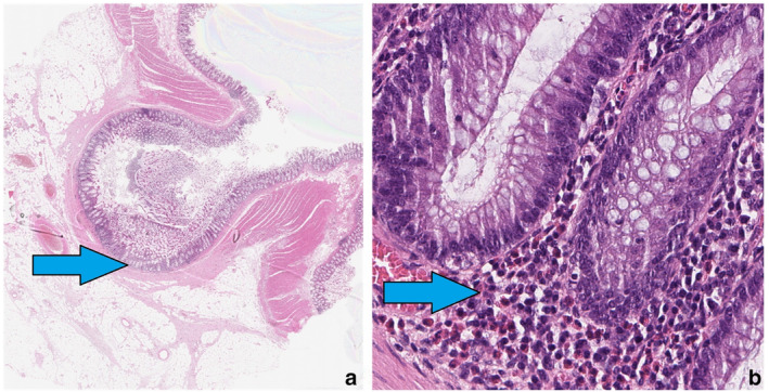
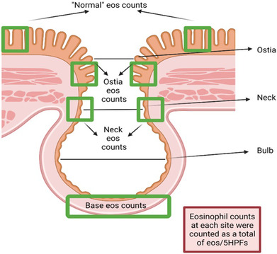
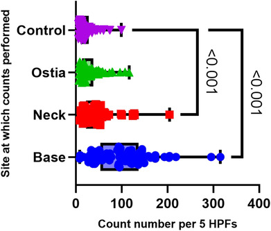
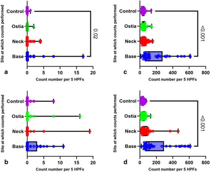
Methods: Hematoxylin and eosin stained sections from colonic surgical resections (n = 82) containing diverticula were examined. Eosinophils, neutrophils, and lymphocytes, in five high power fields in the lamina propria were counted at the base, neck, and ostia of the diverticulum and counts compared to non-diverticula mucosa. The cohort was further subgrouped by elective and emergency surgical indications.
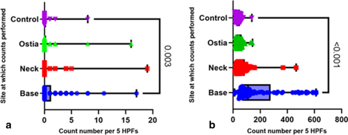
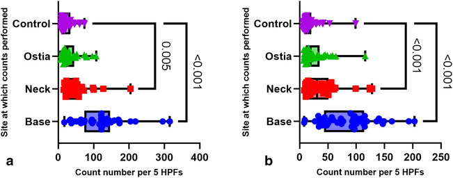
Results: Following an initial review of 10 surgical resections from patients with diverticulosis, a total of 82 patients with colonic resections containing diverticula from the descending colon were evaluated (median age 71.5, 42 M/40F). Eosinophil counts for the entire cohort were increased in the base and neck (median 99 and 42, both P = <0.001) compared with the control location (median 16). Eosinophil counts remained significantly increased in the diverticula base (both P = <0.001) and neck (P = 0.01 and <0.001, respectively) in both elective and emergency cases. Lymphocytes were also significantly increased at the diverticula base compared to controls in both elective and emergency subgroups.
Conclusion: Eosinophils are significantly and most strikingly increased within the diverticulum in resected colonic diverticula. While these observations are novel, the role of eosinophil and chronic inflammation is as yet unclear in the pathophysiology of colonic diverticulosis and diverticular disease.
Keywords: Anatomical histopathology; Diverticular disease; Diverticulitis; Diverticulosis; Eosinophil; Epidemiology; Gastroenterology; Inflammation.
© 2023 The Authors. Journal of Gastroenterology and Hepatology published by Journal of Gastroenterology and Hepatology Foundation and John Wiley & Sons Australia, Ltd.
Figures
Similar articles
-
Surgical management of colonic diverticular disease: discrepancy between right- and left-sided diseases.World J Gastroenterol. 2014 Aug 7;20(29):10115-20. doi: 10.3748/wjg.v20.i29.10115. World J Gastroenterol. 2014. PMID: 25110438 Free PMC article.
-
The pathology of diverticulosis: classical concepts and mucosal changes in diverticula.J Clin Gastroenterol. 2006 Aug;40 Suppl 3:S126-31. doi: 10.1097/01.mcg.0000225508.90417.07. J Clin Gastroenterol. 2006. PMID: 16885695 Review.
-
Diverticulosis and Diverticulitis.Mayo Clin Proc. 2016 Aug;91(8):1094-104. doi: 10.1016/j.mayocp.2016.03.012. Epub 2016 May 5. Mayo Clin Proc. 2016. PMID: 27156370 Review.
-
Colonic Diverticula Are Not Associated With Mucosal Inflammation or Chronic Gastrointestinal Symptoms.Clin Gastroenterol Hepatol. 2018 Jun;16(6):884-891.e1. doi: 10.1016/j.cgh.2017.05.051. Epub 2017 Jun 8. Clin Gastroenterol Hepatol. 2018. PMID: 28603053 Free PMC article.
-
Pathogenesis of colonic diverticulitis and diverticulosis.Postgrad Med. 1976 Dec;60(6):76-81. doi: 10.1080/00325481.1976.11708406. Postgrad Med. 1976. PMID: 792842 Review.
Cited by
-
Beyond Eosinophilic Esophagitis: Eosinophils in Gastrointestinal Disease-New Insights, "New" Diseases.J Can Assoc Gastroenterol. 2023 Nov 24;6(6):199-211. doi: 10.1093/jcag/gwad046. eCollection 2023 Dec. J Can Assoc Gastroenterol. 2023. PMID: 38106480 Free PMC article. Review.
References
-
- Walker MM, Harris AK. Pathogenesis of diverticulosis and diverticular disease. Minerva Gastroenterol. Dietol. 2017; 63: 99–109. - PubMed
-
- Feuerstein JD, Falchuk KR. Diverticulosis and diverticulitis. Mayo Clin. Proc. 2016; 91: 1094–1104. - PubMed
-
- West AB, Losada M. The pathology of diverticulosis coli. J. Clin. Gastroenterol. 2004; 38: S11–S16. - PubMed
-
- West AB, NDSG . The pathology of diverticulitis. J. Clin. Gastroenterol. 2008; 42: 1137–1138. - PubMed
MeSH terms
Grants and funding
LinkOut - more resources
Full Text Sources
Medical
