

Umbilical cord hemangioma and pseudocyst with favorable fetal outcome
- PMID: 37415590
- PMCID: PMC10319952
- DOI: 10.1002/ccr3.7656
Umbilical cord hemangioma and pseudocyst with favorable fetal outcome
Abstract
Key clinical message: There is a high association between umbilical cord hemangiomas or cysts with fetal mortality. However, favorable outcome is possible with proper prenatal monitoring and care.
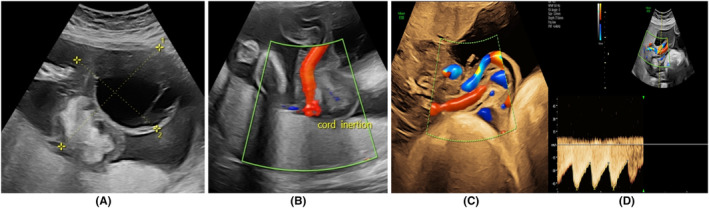
Abstract: Umbilical cord hemangiomas are rare neoplasms of vascular origin, commonly found in the free section of the umbilical cord proximal to placental insertion. They are associated with an increased risk of fetal mortality. We present a rare co-occurrence of an umbilical cord hemangioma and a pseudocyst managed conservatively, with favorable fetal outcome despite the interval increase in size, decreased caliber of the umbilical arteries, and fetal chest compression.
Keywords: fetal outcome; hemangioma; pseudocyst; tumor; umbilical cord.
© 2023 The Authors. Clinical Case Reports published by John Wiley & Sons Ltd.
Conflict of interest statement
The authors have no conflict of interest to declare.
Figures


References
-
- Papadopoulos VG, Kourea HP, Adonakis GL, Decavalas GO. A case of umbilical cord hemangioma: Doppler studies and review of the literature. Eur J Obstet Gynecol Reprod Biol. 2009;144(1):8‐14. - PubMed
-
- Leung KY, Poon CF, Teotico AR, et al. Recommendations on routine mid‐trimester anomaly scan. J of Obstet Gynaecol Res. 2015;41(5):653‐661. - PubMed
Publication types
LinkOut - more resources
Full Text Sources
