. 2023 Jun 21:14:1204045.
doi: 10.3389/fmicb.2023.1204045. eCollection 2023.

The universal Sua5/TsaC family evolved different mechanisms for the synthesis of a key tRNA modification

Affiliations

The universal Sua5/TsaC family evolved different mechanisms for the synthesis of a key tRNA modification

Adeline Pichard-Kostuch et al. Front Microbiol. .

Abstract

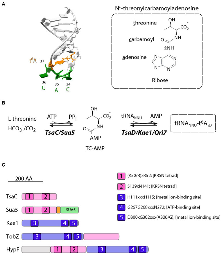
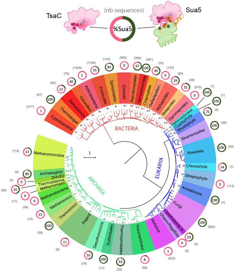
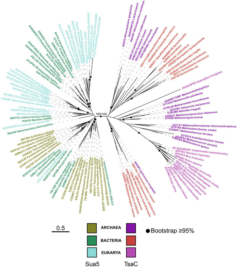
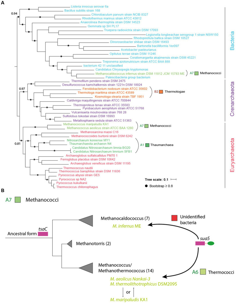
TsaC/Sua5 family of enzymes catalyzes the first step in the synthesis of N6-threonyl-carbamoyl adenosine (t6A) one of few truly ubiquitous tRNA modifications important for translation accuracy. TsaC is a single domain protein while Sua5 proteins contains a TsaC-like domain and an additional SUA5 domain of unknown function. The emergence of these two proteins and their respective mechanisms for t6A synthesis remain poorly understood. Here, we performed phylogenetic and comparative sequence and structure analysis of TsaC and Sua5 proteins. We confirm that this family is ubiquitous but the co-occurrence of both variants in the same organism is rare and unstable. We further find that obligate symbionts are the only organisms lacking sua5 or tsaC genes. The data suggest that Sua5 was the ancestral version of the enzyme while TsaC arose via loss of the SUA5 domain that occurred multiple times in course of evolution. Multiple losses of one of the two variants in combination with horizontal gene transfers along a large range of phylogenetic distances explains the present day patchy distribution of Sua5 and TsaC. The loss of the SUA5 domain triggered adaptive mutations affecting the substrate binding in TsaC proteins. Finally, we identified atypical Sua5 proteins in Archaeoglobi archaea that seem to be in the process of losing the SUA5 domain through progressive gene erosion. Together, our study uncovers the evolutionary path for emergence of these homologous isofunctional enzymes and lays the groundwork for future experimental studies on the function of TsaC/Sua5 proteins in maintaining faithful translation.

Keywords: Sua5; TsaC; enzyme; evolution; t6A; tRNA; universal proteins.


Conflict of interest statement

The authors declare that the research was conducted in the absence of any commercial or financial relationships that could be construed as a potential conflict of interest.

Figures

Figure 1
Figure 1
Structure and biosynthesis of t6A37 nucleoside. (A) On the left is shown the structure of the anticodon loop of Schizosaccharomyces pombe tRNAiMet (PDB 2G1G). The anticodon residues and t6A37 nucleoside are colored in green and orange, respectively. The chemical formula of t6A37 is depicted on the right. (B) Proposed mechanism for t6A37 biosynthesis. (C) Primary structures of proteins containing TsaC orthologous domains. All proteins are drawn to scale and the approximate position of conserved motifs (1–5) is indicated. The numbering corresponds to that of E. coli TsaC and TsaD proteins. Sua5 proteins contain TsaC-orthologous domain with a loop of about 20 residues (orange) fused to SUA5 domain of about 100 residues (green). TobZ proteins catalyse the formation of the antibiotic nebramycin using the hydrolysis of carbamoyl phosphate and its subsequent adenylation by ATP to yield O-carbamoyladenylate. HypF is a carbamoyl transferase involved in the maturation of [NiFe] hydrogenases. HypF uses carbamoylphosphate as a substrate and transfers the carboxamido moiety in an ATP-dependent reaction to the thiolate of the C-terminal cysteine of HypE yielding a protein-S-carboxamide.
Figure 2
Figure 2
Distribution of TsaC and Sua5 proteins across the tree of life. The universal phylogeny based on RNA polymerase sequences is depicted. The number of TsaC and Sua5 sequences is indicated in the brackets for each taxon. The ring graph indicates the ratio of TsaC (pink) and Sua5 (green) orthologs for a given taxon. The numbers in the ring correspond to the percentage of Sua5 sequences. The scale bar corresponds to the number of substitutions per position in the alignment.
Figure 3
Figure 3
The phylogeny of the TsaC/Sua5 family of proteins. The maximum likelihood tree was generated from the alignment of representative TsaC and Sua5 sequences (see Supplementary Tables 2,3) using LG + R6 sequence evolution model. Sequences of TsaC and TsaC-like domain of Sua5 proteins were used for the alignment. The scale bar corresponds to the number of substitutions per amino acid in the alignment.
Figure 4
Figure 4
Horizontal transfer of sua5 genes occurs across different phylogenetic distances. (A) Maximum likelihood phylogenetic tree of Sua5 sequences from a wide range of bacterial taxa and from representative sequences of Crenarchaeota and Euryarchaeaota. The tree was arbitrarily rooted between Bacteria and Archaea. The Sua5 sequences from Methanococci, Thaumarchaea and Thermotogae are indicated in colors corresponding to their taxonomic group (see Supplementary Table 1). The scale bar corresponds to the number of substitutions per amino acid. (B) Evolutionary scenario for the acquisition of sua5 genes in Methanococci archaea. The established phylogeny of the four main genera of Methanococci is shown. The number of species in each genus is indicated in brackets. All species carry TsaC orthologs except for the four species (indicated in green color) that carry Sua5 orthologs. This suggest that the common ancestor of Methanococci was a TsaC-user and that the four species acquired the sua5 gene by HGT. The putative donor of these sua5 genes is indicated above the arrows.
Figure 5
Figure 5
Variant-specific conserved residues interact with substrates and stabilize interdomain interface. (A) Cartoon showing the structure of Pa-Sua5 in complex with threonine and PPi shown as stick presentation. The TsaC-like domain is in pink, SUA5 domain is in green and the flexible loop is in orange. The Sua5-specific conserved residues are indicated with one letter code in pink circles or in cyan circle for the signature residue. (B) Cartoon showing the crystal structure of Ec-TsaC. Threonine and PPi molecules were modelled in the active site by superposing the Ec-TsaC with Pa-Sua5. The signature residue Thr is indicated with one code letter in a cyan-colored circle. (C) Zoom in the active site of Pa-Sua5. Sua5-specific conserved residues Pro59, Asn62, His67, and Pro142 are highlighted. Asn62 forms a hydrogen bond (indicated as dotted line) with the PPi molecule. (D) Zoom at the interface between the TsaC-like and SUA5 domains. The KPSPT motif and the GVE motif are shown as well as the network of H-bonds formed between these residues. Two salt bridges formed by Asp161 and Glu180 with Arg301 and Arg328, respectively, likely stabilize the two domains. (E) Zoom in the active site cavity of Ec-TsaC. Threonine and PPi molecules were modelled in the active site by superposing the Ec-TsaC and Pa-Sua5 that co-crystallized with these ligands. Side chain of the signature residue Thr138 is shown as stick model. (F) Zoom in the active site of TsaC-like domain of TobZ protein from Streptoalloteichues tenebrarius bound to its substrate carbamoyl-phosphate (CP). The side chain of Thr529 (the equivalent of Thr138 in Ec-TsaC) is indicated as stick model. The H-bond between Thr529 and the phosphate moiety is indicated by a dotted line.
Figure 6
Figure 6
Nature and frequency of the signature residue in TsaC and Sua5 sequences Nature of the signature residue Pro143/Thr138/Ser138 in TsaC and Sua5 sequences from Eukaryotes (A) and Prokaryotes (B). The top graph shows the protein length distribution whereby each sequence is represented as a vertical bar. The number of analyzed sequences is indicated under the graph. The legend gives the correspondence between the color of the bars and the presence of one of the signature residues. Bottom cartoons depict the percentage of TsaC and Sua5 having either Pro143, Thr138, or Ser138as the signature residue.
Figure 7
Figure 7
Progressive erosion of the SUA5 domain in Sua5 proteins from Archaeoglobus archaea. (A) Maximum likelihood tree of Sua5 proteins from Archaeoglobi species. The isolated species are highlighted in blue color. Bootstrap values for branch support are indicated. The table on the right shows the occurrence of the Sua5-specific conserved residues in the analyzed sequences. The canonical residues are indicated on the top of the table. The residues found in Ap-Sua5 are highlighted in grey and signature residue Pro143/Thr138 is indicated in cyan. Residues diverging from the consensus sequence and indels are in orange. (B) AlphaFold2 model of Ap-Sua5 (in color) is superposed onto the crystal structure of Pa-Sua5 shown in light gray. The consensus Sua5-specific residues and the corresponding residues found in Ap-Sua5 are indicated as spheres. (C) Structure of Sua5 proteins shown as molecular surface. The crystal structure of Pa-Sua5 was retrieved from the PDB database while the structures of Av-Sua5, Af-Sua5, and Ap-Sua5 were modeled using AlphaFold2. TsaC-like domain, the interdomain loop, and SUA5 domain are depicted in pink, orange, and green color, respectively.

Similar articles

Cited by

References

    1. Agari Y., Sato S., Wakamatsu T., Bessho Y., Ebihara A., Yokoyama S., et al. . (2008). X-ray crystal structure of a hypothetical Sua5 protein from Sulfolobus tokodaii strain 7. Proteins Struct. Funct. Bioinform. 70, 1108–1111. doi: 10.1002/prot.21794 - DOI - PubMed
    1. Arrondel C., Missoury S., Snoek R., Patat J., Menara G., Collinet B., et al. . (2019). Defects in t(6)a tRNA modification due to GON7 and YRDC mutations lead to Galloway-Mowat syndrome. Nat. Commun. 10:3967. doi: 10.1038/s41467-019-11951-x - DOI - PMC - PubMed
    1. Baldauf S. L., Palmer J. D., Doolittle W. F. (1996). The root of the universal tree and the origin of eukaryotes based on elongation factor phylogeny. Proc. Natl. Acad. Sci. U.S. A. 93, 7749–7754. doi: 10.1073/pnas.93.15.7749 - DOI - PMC - PubMed
    1. Beenstock J., Sicheri F. (2021). The structural and functional workings of KEOPS. Nucleic Acids Res. 49, 10818–10834. doi: 10.1093/nar/gkab865 - DOI - PMC - PubMed
    1. Bornberg-Bauer E., Huylmans A.-K., Sikosek T. (2010). How do new proteins arise? Curr. Opin. Struct. Biol. 20, 390–396. doi: 10.1016/j.sbi.2010.02.005 - DOI - PubMed

LinkOut - more resources