Tinea pedis: an updated review
- PMID: 37415917
- PMCID: PMC10321471
- DOI: 10.7573/dic.2023-5-1
Tinea pedis: an updated review
Abstract
Background: Tinea pedis is one of the most common superficial fungal infections of the skin, with various clinical manifestations. This review aims to familiarize physicians with the clinical features, diagnosis and management of tinea pedis.
Methods: A search was conducted in April 2023 in PubMed Clinical Queries using the key terms 'tinea pedis' OR 'athlete's foot'. The search strategy included all clinical trials, observational studies and reviews published in English within the past 10 years.
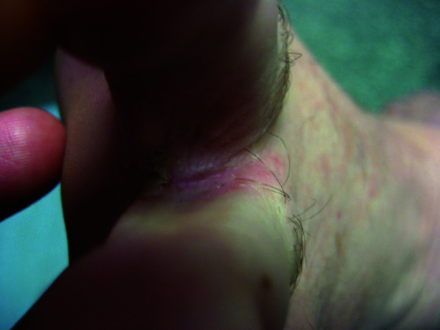
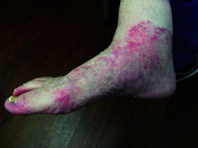
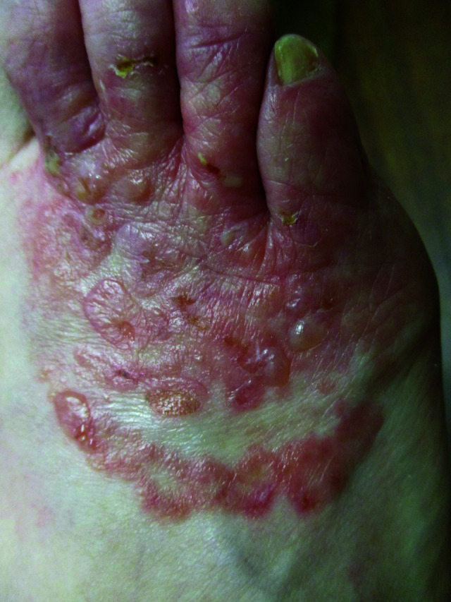
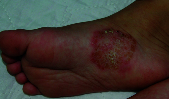
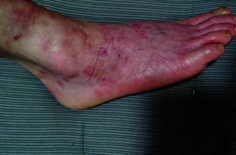
Results: Tinea pedis is most often caused by Trichophyton rubrum and Trichophyton interdigitale. It is estimated that approximately 3% of the world population have tinea pedis. The prevalence is higher in adolescents and adults than in children. The peak age incidence is between 16 and 45 years of age. Tinea pedis is more common amongst males than females. Transmission amongst family members is the most common route, and transmission can also occur through indirect contact with contaminated belongings of the affected patient. Three main clinical forms of tinea pedis are recognized: interdigital, hyperkeratotic (moccasin-type) and vesiculobullous (inflammatory). The accuracy of clinical diagnosis of tinea pedis is low. A KOH wet-mount examination of skin scrapings of the active border of the lesion is recommended as a point-of-care testing. The diagnosis can be confirmed, if necessary, by fungal culture or culture-independent molecular tools of skin scrapings. Superficial or localized tinea pedis usually responds to topical antifungal therapy. Oral antifungal therapy should be reserved for severe disease, failed topical antifungal therapy, concomitant presence of onychomycosis or in immunocompromised patients.
Conclusion: Topical antifungal therapy (once to twice daily for 1-6 weeks) is the mainstay of treatment for superficial or localized tinea pedis. Examples of topical antifungal agents include allylamines (e.g. terbinafine), azoles (e.g. ketoconazole), benzylamine, ciclopirox, tolnaftate and amorolfine. Oral antifungal agents used for the treatment of tinea pedis include terbinafine, itraconazole and fluconazole. Combined therapy with topical and oral antifungals may increase the cure rate. The prognosis is good with appropriate antifungal treatment. Untreated, the lesions may persist and progress.
Keywords: Trichophyton interdigitale; Trichophyton rubrum; athlete’s foot; dermatophytosis; interdigital; moccasin; vesiculobullous.
Copyright © 2023 Leung AKC, Barankin B, Lam JM, Leong KF, Hon KL.
Conflict of interest statement
Disclosure and potential conflicts of interest: AKCL and KLH are associate editors of Drugs in Context and confirm that this article has no other conflicts of interest otherwise. This manuscript was sent out for independent peer review. The International Committee of Medical Journal Editors (ICMJE) Potential Conflicts of Interests form for the authors is available for download at: https://www.drugsincontext.com/wp-content/uploads/2023/06/dic.2023-5-1-COI.pdf
Figures

References
-
- Kaštelan M, Utješinović-Gudelj V, Prpić-Massari L, Brajac I. Dermatophyte infections in Primorsko-Goranska County, Croatia: a 21-year survey. Acta Dermatovenerol Croat. 2014;22(3):175–179. - PubMed
Publication types
LinkOut - more resources
Full Text Sources
Miscellaneous