

Glycan dependent phenotype differences of HIV-1 generated from macrophage versus CD4+ T helper cell populations
- PMID: 37415979
- PMCID: PMC10320205
- DOI: 10.3389/fimmu.2023.1107349
Glycan dependent phenotype differences of HIV-1 generated from macrophage versus CD4+ T helper cell populations
Abstract
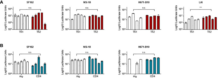
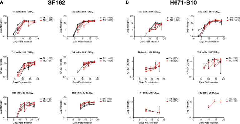
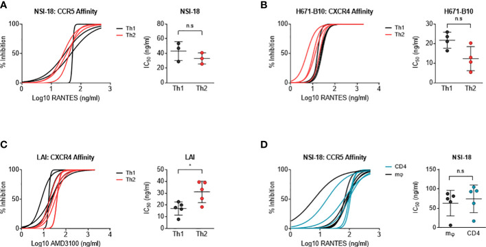
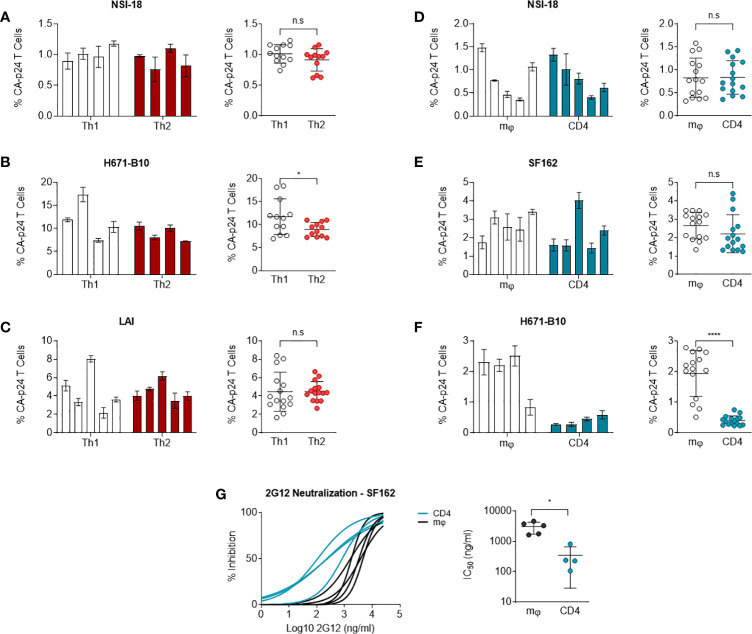
Human immunodeficiency virus type 1 (HIV-1) is able to infect a variety of cell types with differences in entry efficiency and replication kinetics determined by the host cell type or the viral phenotype. The phenotype of the virus produced from these various cell types, including infectivity, co-receptor usage and neutralisation sensitivity, may also be affected by the characteristics of the producing cell. This can be due to incorporation of variant cell-specific molecules or differences in post-translational modifications of the gp41/120 envelope. In this study we produced genetically identical virus strains from macrophages, CD4-enriched lymphocytes as well as Th1 and Th2 CD4+ cell lines and compared each different virus stock for their infectivity in various cell types and sensitivity to neutralisation. In order to study the effect of the producer host cell on the virus phenotype, virus stocks were normalised on infectivity and were sequenced to confirm env gene homogeneity. Virus production by Th1 or Th2 cells did not compromise infectivity of the variant cell types tested. We observed no difference in sensitivity to co-receptor blocking agents upon viral passage through Th1 and Th2 CD4+ cell lineages nor did this affect DC-SIGN-mediated viral capture as measured in a transfer assay to CD4+ lymphocytes. Virus produced by macrophages was comparably sensitive to CC-chemokine inhibition as was virus generated from the array of CD4+ lymphocytes. We identified that virus produced from macrophages was fourteen times more resistant to 2G12 neutralisation than virus produced from CD4+ lymphocytes. Macrophage-produced dual-tropic (R5/X4) virus was six times more efficiently transmitted to CD4+ cells than lymphocyte-derived HIV-1 (p<0.0001) after DCSIGN capture. These results provide further insights to what extent the host cell influences viral phenotype and thereby various aspects of HIV-1 pathogenesis but suggest that viruses generated from Th1 versus Th2 cells are consistent in phenotype.
Keywords: HIV-1; T helper; glycan; macrophages; phenotypes.
Copyright © 2023 Heeregrave, Thomas, van Capel, de Jong, Pollakis and Paxton.
Conflict of interest statement
The authors declare that the research was conducted in the absence of any commercial or financial relationships that could be construed as a potential conflict of interest.
Figures
Similar articles
-
Phenotypic Correlates of HIV-1 Macrophage Tropism.J Virol. 2015 Nov;89(22):11294-311. doi: 10.1128/JVI.00946-15. Epub 2015 Sep 2. J Virol. 2015. PMID: 26339058 Free PMC article.
-
Envelope-dependent restriction of human immunodeficiency virus type 1 spreading in CD4(+) T lymphocytes: R5 but not X4 viruses replicate in the absence of T-cell receptor restimulation.J Virol. 1999 Sep;73(9):7515-23. doi: 10.1128/JVI.73.9.7515-7523.1999. J Virol. 1999. PMID: 10438841 Free PMC article.
-
Quantification of entry phenotypes of macrophage-tropic HIV-1 across a wide range of CD4 densities.J Virol. 2014 Feb;88(4):1858-69. doi: 10.1128/JVI.02477-13. Epub 2013 Dec 4. J Virol. 2014. PMID: 24307580 Free PMC article.
-
HIV-1 replication in CD4+ T cell lines: the effects of adaptation on co-receptor use, tropism, and accessory gene function.J Leukoc Biol. 2000 Sep;68(3):331-7. J Leukoc Biol. 2000. PMID: 10985248 Review.
-
Evolution of Host Target Cell Specificity During HIV-1 Infection.Curr HIV Res. 2018;16(1):13-20. doi: 10.2174/1570162X16666171222105721. Curr HIV Res. 2018. PMID: 29268687 Review.
Cited by
-
Macrophage- and CD4+ T cell-derived SIV differ in glycosylation, infectivity and neutralization sensitivity.PLoS Pathog. 2024 May 28;20(5):e1012190. doi: 10.1371/journal.ppat.1012190. eCollection 2024 May. PLoS Pathog. 2024. PMID: 38805549 Free PMC article.
-
Glycan heterogeneity as a cause of the persistent fraction in HIV-1 neutralization.PLoS Pathog. 2023 Oct 30;19(10):e1011601. doi: 10.1371/journal.ppat.1011601. eCollection 2023 Oct. PLoS Pathog. 2023. PMID: 37903160 Free PMC article.
References
-
- Brenchley JM, Hill BJ, Ambrozak DR, Price DA, Guenaga FJ, Casazza JP, et al. . T-Cell subsets that harbor human immunodeficiency virus (HIV) In vivo: implications for HIV pathogenesis. J Virol (2004) 78:1160–8. doi: 10.1128/JVI.78.3.1160-1168.2004/ASSET/89741904-EED1-448C-8AB9-DBF28EA4229B/ASSETS/GRAPHIC/ZJV0030416010007.JPEG - DOI - PMC - PubMed
-
- Dornadula G, Zhang H, Shetty S, Pomerantz RJ. HIV-1 virions produced from replicating peripheral blood lymphocytes are more infectious than those from nonproliferating macrophages due to higher levels of intravirion reverse transcripts: implications for pathogenesis and transmission. Virology (1999) 253:10–6. doi: 10.1006/VIRO.1998.9465 - DOI - PubMed
Publication types
MeSH terms
Substances
LinkOut - more resources
Full Text Sources
Research Materials