. 2023 Jun 15;26(7):107142.
doi: 10.1016/j.isci.2023.107142. eCollection 2023 Jul 21.

High-throughput longitudinal electrophysiology screening of mature chamber-specific hiPSC-CMs using optical mapping

Affiliations

High-throughput longitudinal electrophysiology screening of mature chamber-specific hiPSC-CMs using optical mapping

Andrew Allan et al. iScience. .

Abstract

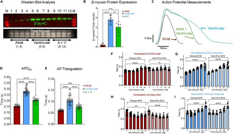
hiPSC-CMs are being considered by the Food and Drug Administration and other regulatory agencies for in vitro cardiotoxicity screening to provide human-relevant safety data. Widespread adoption of hiPSC-CMs in regulatory and academic science is limited by the immature, fetal-like phenotype of the cells. Here, to advance the maturation state of hiPSC-CMs, we developed and validated a human perinatal stem cell-derived extracellular matrix coating applied to high-throughput cell culture plates. We also present and validate a cardiac optical mapping device designed for high-throughput functional assessment of mature hiPSC-CM action potentials using voltage-sensitive dye and calcium transients using calcium-sensitive dyes or genetically encoded calcium indicators (GECI, GCaMP6). We utilize the optical mapping device to provide new biological insight into mature chamber-specific hiPSC-CMs, responsiveness to cardioactive drugs, the effect of GCaMP6 genetic variants on electrophysiological function, and the effect of daily β-receptor stimulation on hiPSC-CM monolayer function and SERCA2a expression.

Keywords: Cell biology; Medicine; Screening in health technology.


Conflict of interest statement

T.J.H. is a consultant to StemBioSys, Inc. and is a member of the Scientific Advisory Board. A.M.R., J.C., T.J.H., and T.B. have ownership stake in StemBioSys, Inc. T.B. is an employee of StemBioSys, Inc. A.A. is an employee of CAIRN Research. D.-H.K. is a scientific founder and equity holder of Curi Bio.

Figures

None
Graphical abstract
Figure 1
Figure 1
MatrixPlus-human ECM promotes maturation of hiPSC-CM structure and mitochondrial function (A) hiPSC-CM mitochondrial function was measured using JC-1 mitochondrial-specific fluorescent marker. (B) JC-1 red-to-green ratio was greater in hiPSC-CMs plated on MatrixPlus-human ECM (4.51 ± 1.69 fluorescence units, n = 102 hiPSC-CMs) than hiPSC-CMs plated on Matrigel-mouse ECM (3.90 ± 1.43 fluorescence units, n = 114 hiPSC-CMs). ∗∗p = 0.0046, unpaired t test. (C) Sarcomere staining using α-actinin-specific antibody and confocal imaging shows distinct sarcomere organization between hiPSC-CMs plated on Matrigel-mouse ECM and MatrixPlus-human ECM. The white arrow in each picture was used to make the intensity profile plots for α-actinin periodicity over 25μm. (D) hiPSC-CMs maintained on Matrigel-mouse ECM have significantly shorter sarcomere length (1.61 ± 0.17μm, n = 129) than hiPSC-CMs maintained on MatrixPlus-human ECM (1.86 ± 0.14μm, n = 129). ∗∗∗∗p < 0.0001, unpaired t test. (E) Live cell phase-contrast images (20X) show the difference of shape between hiPSC-CMs maintained on Matrigel-mouse ECM compared to the same batch of hiPSC-CMs maintained on MatrixPlus-human ECM. (F) hiPSC-CM circularity index was quantified using multiple hiPSC lines with chamber specification or without chamber specification. For each cell line, black symbols represent the cell circularity on Matrigel-mouse ECM while red symbols represent the cell circularity on MatrixPlus-human ECM. Statistical comparisons were made within each group plated on each ECM. ∗∗∗∗p < 0.0001, unpaired t tests, n = 77–109 hiPSC-CMs per group. (G) Optical mapping calcium transient wave conduction was faster in hiPSC-CM monolayers maintained on MatrixPlus-human ECM (Matrigel = 21.2 ± 4.0 cm/s; n = 7 vs. MatrixPlus-human ECM = 29.8 ± 5.9 cm/s; n = 7. ∗∗p = 0.0078, unpaired t test. Data are expressed as mean ± standard deviation.
Figure 2
Figure 2
Validation of voltage-sensitive dye and calcium-sensitive dye high-throughput electrophysiology assays (A) Representative recording of atrial hiPSC-CM monolayer action potentials using FluoVolt. (B) Representative recording of atrial hiPSC-CM monolayer calcium transients using CalBryte 520AM. (C) The spontaneous beat rate of atrial hiPSC-CM monolayers was similar regardless of parameter measured (Vm = 0.98 ± 0.19 Hz, n = 56 monolayers; CaT = 0.91 ± 0.21 Hz, n = 32 monolayers; unpaired t test, ns). This represents 56 and 32 technical monolayer replicates, respectively. (D) Impulse conduction velocity was similar between each parameter measured (Vm = 46.7 ± 7.5 cm s-1, n = 55 monolayers; CaT = 44.5 ± 11.8 cm s-1, n = 32 monolayers; unpaired t test, ns). (E) APD80 was significantly shorter than CaTD80 (Vm = 0.170 ± 0.04 s, n = 56 monolayers; CaT = 0.289 ± 0.05 s, n = 32 monolayers technical replicates; unpaired t test, ∗∗∗∗p < 0.0001). (F) AP triangulation was significantly lower than CaT triangulation (Vm = 0.06 ± 0.01 s, n = 56 monolayers; CaT = 0.22 ± 0.04 s, n = 32 monolayers; unpaired t test, ∗∗∗∗p < 0.0001). (G) APD or CaT duration 30 heatmap for 8 representative wells per group. (H) APD or CaT duration 90 heatmap for 8 representative wells per group. (I) AP or CaT triangulation heatmap for 8 representative wells per group. Data are expressed as mean ± standard deviation.
Figure 3
Figure 3
Cardiac optical mapping validation using three cardioactive compounds applied to atrial hiPSC-CM monolayers (A) Representative before and after calcium transient traces of a single well treated with isoproterenol (+ISO, 0.10μM). (B) Paired analysis indicates increased spontaneous beat rate after ISO treatment. (Paired t test, ∗∗∗p = 0.002, n = 20 monolayer technical replicates). (C) Impulse conduction velocity was increased following ISO treatment. (Paired t test, ∗∗∗∗p < 0.0001, n = 20 monolayers). (D) Calcium transient amplitude (ΔF/F0) was increased after ISO treatment. (Paired t test, ∗∗∗∗p < 0.0001). (E) CaTD80 was slightly greater following ISO treatment. (Paired t test p < 0.0001). (F) Representative before and after calcium transient traces of a single monolayer treated with E−4031 (+E−4031, 0.5μM). (G) Paired analysis indicates lack of effect of E−4031 on atrial hiPSC-CM monolayer beat rate. (H) Conduction velocity was significantly slower with E−4031 treatment. (Paired t test, ∗∗∗∗p < 0.0001, n = 20 monolayers). (I) Calcium transient amplitude (ΔF/F0) was reduced by E−4031 treatment. (Paired t test, ∗∗∗∗p < 0.0001). (J) E−4031 significantly increased CaTD80. (Paired t test, ∗∗∗∗p < 0.0001). (K) Representative traces show the effect of Ibutilide (0.1μM) on the calcium transient. (L) Ibutilide slightly increased the beat rate. (Paired t test, ∗∗p = 0.007). (M) Ibutilide slowed conduction velocity. (Paired t test, ∗∗∗∗p < 0.0001, n = 20 monolayer technical replicates). (N) Ibutilide treatment reduced the calcium transient amplitude (ΔF/F0). (Paired t test, ∗∗∗∗p < 0.0001). (O) Ibutilide significantly increased CaTD80. (Paired t test, ∗∗∗∗p < 0.0001). Data are expressed as mean ± standard deviation.
Figure 4
Figure 4
High-throughput electrophysiology analysis of chamber-specific hiPSC-CMs (A) Western blot analysis of β-myosin heavy chain (β-MyHC) isoform expression in purified atrial-iPSC-CMs and purified ventricular-iPSC-CMs. (B) Quantification of β-MyHC relative to total myosin indicates that ventricular hiPSC-CMs express significantly greater β-MyHC (2.81 ± 0.44au, n = 4 monolayer technical replicates) than atrial hiPSC-CMs (0.21 ± 0.16au, n = 4 monolayers). (One-way ANOVA, ∗∗∗∗p < 0.0001; ∗∗∗p = 0.0005). (C and D) Optical mapping in 96-well plates, using voltage-sensitive dye (VSD, FluoVolt) confirms that atrial hiPSC-CM monolayers have shorter action potential duration (0.154 ± 0.01s, n = 18 monolayer technical replicates) than ventricular hiPSC-CMs (0.281 ± 0.03s, n = 20 monolayers) and monolayers made of 50% atrial and 50% ventricular hiPSC-CMs had average APD80 in between the two extremes (0.207 ± 0.02s, n = 20 monolayers). (One-way ANOVA, ∗∗∗∗p < 0.0001). (E) AP triangulation is greater in the ventricular monolayers (0.104 ± 0.02, n = 20 monolayers) than in atrial monolayers (0.060 ± 0.004, n = 18 monolayers) and heterogeneous monolayers of 50% atrial and 50% ventricular (A + V, 0.077 ± 0.008, n = 20 monolayers). (One-way ANOVA, ∗∗∗∗p < 0.0001; ∗∗∗p = 0.0006). For ANOVA post hoc analysis, Tukey’s multiple comparisons test was used. (F) Vernakalant dose response for effect on atrial CM CaTD30 plated on Matrigel (solid bars) or MatrixPlus (hashed bars).∗∗∗∗p < 0.0001, unpaired t test 0 vernakalant vs. 30μM. (G) Vernakalant dose response for the effect on ventricular CM CaTD30 plated on Matrigel (solid bars) or MatrixPlus (hashed bars). ∗∗∗∗p < 0.0001, unpaired t test 0 vernakalant vs. 30μM. (H) Dose response of vernakalant effect on Ca Triangulation (Ca Triang) in atrial CMs plated on Matrigel (solid bars) or MatrixPlus (hashed bars). ∗∗∗∗p < 0.0001, ∗∗p = 0.008, unpaired t test 0 vernakalant vs. 30μM. (I) Dose response of vernakalant effect on Ca Triangulation (Ca Triang) in ventricular CMs plated on Matrigel (solid bars) or MatrixPlus (hashed bars). ∗∗∗∗p < 0.0001, unpaired t test, 0 vernakalant vs. 30μM. Data are expressed as mean ± standard deviation.
Figure 5
Figure 5
Characterization of GCaMP6 variants effects on hiPSC-CM electrophysiological function with repeated measures (A) Representative calcium transient traces recorded on day 2 following AdGCaMP6f, m, or s viral transduction in commercially available hiPSC-CMs (iCell, Cellular Dynamics International) using a widefield fluorescence microscope. (B) Day 2 microscopic images of monolayers’ baseline green fluorescence for each GCaMP6 variant: red = fast, blue = medium and green = slow. (C) Calcium transients recorded on day 5 following adenoviral transduction of each GCaMP6 variant. (D) Day 5 microscopic images of monolayers’ baseline green fluorescence for each GCaMP6 variant. (E) Calcium transients recorded on day 7 following adenoviral transduction of each GCaMP6 variant. (F) Day 7 microscopic images of monolayers’ baseline green fluorescence for each GCaMP6 variant. (G–I) Representative CARTOX optical recordings of hiPSC-CM calcium transients on day 3 after adenoviral transduction of each GCaMP6 variant. (J–L) Day 4 representative recordings of calcium transients and M-O show Day 7 recordings of each GCaMP6 variant. (P–S) Quantification of calcium transient duration 80 (CaTD80) for each variant at days 3–7. (One-way ANOVA, ∗∗∗∗p < 0.0001, ∗∗∗p = 0.0002, ∗∗p < 0.008, ∗p < 0.05, n = 24 monolayer technical replicates per group). (T–W) Quantification of hiPSC-CM spontaneous beat rate days 3–7 following adenoviral transduction of each GCaMP6 variant. (One-way ANOVA, ∗∗∗∗p < 0.0001, ∗∗∗p = 0.0002, ∗∗p < 0.008, ∗p < 0.05, n = 24 monolayers per group). For ANOVA post hoc analysis, Tukey’s multiple comparisons test was used. Data are expressed as mean ± standard deviation.
Figure 6
Figure 6
AdGCaMP6 variant effect on hiPSC-CM responsiveness to two classical cardioactive compounds on day 6 following viral transduction (A–C) representative well traces of calcium transient responses to isoproterenol (+ISO, 0.10μM) stimulation. –ISO indicates traces before treatment +ISO indicates the same well traces immediately following ISO treatment for each GCaMP6 variant (A, red = AdGCaMP6f; B, blue = AdGCaMP6; C, green = AdGCaMP6s. (D) Paired analysis of the spontaneous beat rate for each virus treatment before and after ISO. (E) Paired analysis for the effect of ISO on the calcium transient amplitude (ΔF/F0) in each virus-treated group. (F) Paired analysis for the effect of ISO on the CaTD80 in each virus-treated group. (For D-F; Paired t tests, ∗∗∗∗p < 0.0001; ∗∗∗p ≤ 0.002, n = 23–24 monolayer technical replicates per group). (G–I) Representative well traces of calcium transient responses to E−4031 (0.2μM) for each GCaMP6 variant. (J) Paired analysis of the effect of E−4031 on hiPSC-CM spontaneous beat rate. (K) Paired analysis of the effect of E−4031 on the calcium transient amplitude for each virus-treated group. (L) Paired analysis for the effect of E−4031 on the CaTD80 for each group. (For J-L, Paired t tests, ∗∗∗∗p < 0.0001, ∗∗∗p ≤ 0.002, n = 23–24 monolayers per group). Data are expressed as mean ± standard deviation.
Figure 7
Figure 7
GCaMP6f repeated measures to determine effect of daily isoproterenol pulses on hiPSC-CM function (A) Daily measurement of GCaMP6f spontaneous calcium flux in hiPSC-CM monolayers confirmed pulsing of isoproterenol (0.2μM). At each time point, isoproterenol-treated monolayers’ spontaneous beat rate increased significantly (red symbols, n = 21; ∗∗∗∗p < 0.0001, unpaired t test compared to control, vehicle treated, n = 8, black symbols). (B) On day 5 of this protocol baseline recordings of spontaneous activity was recorded using the cardiac EP plate reader; these are representative traces from each group. Upstroke of the calcium transient is automatically determined and colored green while the calcium transient decay from peak is labeled red. (C) Isoproterenol pulsing increased spontaneous beat rate (Pulsed ISO = 0.65 ± 0.12Hz, n = 8 vs. Control (Vehicle) = 0.46 ± 0.09Hz, n = 16 monolayer technical replicates; ∗∗∗p = 0.0006, unpaired t test). (D) CaTD80 was shorter in ISO-pulsed monolayers (0.623 ± 0.11s, n = 16) relative to control (0.809 ± 0.02s, n = 8); ∗∗∗p = 0.0006, unpaired t test. (E) Calcium transient triangulation was significantly less in ISO-pulsed monolayers (0.309 ± 0.07s, n = 16) compared to control (0.467 ± 0.02s, n = 8) ∗∗∗∗p < 0.0001, unpaired t test. (F) Calcium transient amplitude was not different at baseline between the two groups. (G) Conduction velocity was greater in monolayers pulsed daily with ISO (26.4 ± 5.3 cm/s, n = 16 vs. 20.1 ± 4.9 cm/s, n = 8; ∗∗p = 0.009, unpaired t test). (H) Calcium transient upstroke slope (+dF/dt) was faster in ISO-pulsed monolayers. (I) Western blot analysis probing for SERCA2a expression (top) and phospho-cTnI (middle). Total protein stain (bottom) used for normalization and equal protein loading control. (J) SERCA2a protein expression on day 5 was greater in monolayers treated daily with ISO. ∗∗p = 0.001, unpaired t test, n = 4 per group. (K) In response to acute ISO on day 5, pulsed monolayers beat rate increased to a greater value than control (83.3 ± 13.8 beat per minute (BPM), n = 8 vs. 56.67 ± 20.0BPM, n = 4). (L) Conduction velocity increased to greater values following acute ISO treatment in monolayers with a history of ISO pulsing (48.9 ± 13.2 cm/s, n = 8 vs. 30.1 ± 8.4 cm/s, n = 4 technical replicates) ∗p = 0.02, unpaired t tests. Data are expressed as mean ± standard deviation.

References

    1. Zhang J., Wilson G.F., Soerens A.G., Koonce C.H., Yu J., Palecek S.P., Thomson J.A., Kamp T.J. Functional cardiomyocytes derived from human induced pluripotent stem cells. Circ. Res. 2009;104:e30–e41. - PMC - PubMed
    1. Thomson J.A., Itskovitz-Eldor J., Shapiro S.S., Waknitz M.A., Swiergiel J.J., Marshall V.S., Jones J.M. Embryonic stem cell lines derived from human blastocysts. Science. 1998;282:1145–1147. - PubMed
    1. Takahashi K., Tanabe K., Ohnuki M., Narita M., Ichisaka T., Tomoda K., Yamanaka S. Induction of pluripotent stem cells from adult human fibroblasts by defined factors. Cell. 2007;131:861–872. - PubMed
    1. Ahmed R.E., Anzai T., Chanthra N., Uosaki H. A Brief Review of Current Maturation Methods for Human Induced Pluripotent Stem Cells-Derived Cardiomyocytes. Front. Cell Dev. Biol. 2020;8:178. - PMC - PubMed
    1. Blinova K., Dang Q., Millard D., Smith G., Pierson J., Guo L., Brock M., Lu H.R., Kraushaar U., Zeng H., et al. International Multisite Study of Human-Induced Pluripotent Stem Cell-Derived Cardiomyocytes for Drug Proarrhythmic Potential Assessment. Cell Rep. 2018;24:3582–3592. - PMC - PubMed

LinkOut - more resources