. 2023 Jun 21:17:1212097.
doi: 10.3389/fncel.2023.1212097. eCollection 2023.

A simple method for poly-D-lysine coating to enhance adhesion and maturation of primary cortical neuron cultures in vitro

Affiliations

A simple method for poly-D-lysine coating to enhance adhesion and maturation of primary cortical neuron cultures in vitro

Aurélie Stil et al. Front Cell Neurosci. .

Abstract

Introduction: Glass coverslips are used as a substrate since Harrison's initial nerve cell culture experiments in 1910. In 1974, the first study of brain cells seeded onto polylysine (PL) coated substrate was published. Usually, neurons adhere quickly to PL coating. However, maintaining cortical neurons in culture on PL coating for a prolonged time is challenging.

Methods: A collaborative study between chemical engineers and neurobiologists was conducted to find a simple method to enhance neuronal maturation on poly-D-lysine (PDL). In this work, a simple protocol to coat PDL efficiently on coverslips is presented, characterized, and compared to a conventional adsorption method. We studied the adhesion and maturation of primary cortical neurons with various morphological and functional approaches, including phase contrast microscopy, immunocytochemistry, scanning electron microscopy, patch clamp recordings, and calcium imaging.

Results: We observed that several parameters of neuronal maturation are influenced by the substrate: neurons develop more dense and extended networks and synaptic activity is enhanced, when seeded on covalently bound PDL compared to adsorbed PDL.

Discussion: Hence, we established reproducible and optimal conditions enhancing maturation of primary cortical neurons in vitro. Our method allows higher reliability and yield of results and could also be profitable for laboratories using PL with other cell types.

Keywords: glass coverslip; neuronal adhesion; neuronal maturation; neuronal networks; poly-D-lysine grafting; primary cortical neuron cultures; synaptic activity; synaptic contacts.


Conflict of interest statement

The authors declare that the research was conducted in the absence of any commercial or financial relationships that could be construed as a potential conflict of interest.

Figures

FIGURE 1
FIGURE 1
Simple method of PDL9 covalent binding on glass coverslip. (A) Schematic illustration of the chemical grafting procedures. The grafting of GOPS on glass surfaces is followed by chemical immobilization of PDL chains. (B) Timeline and duration of the different steps from bath base coverslip cleaning to neuron seeding. (C) All steps (base bath, washes, drying, silane deposit, heat drying, PDL grafting and EtA deposit) are illustrated.
FIGURE 2
FIGURE 2
The extracellular matrix of PDL6 is responsible for neuron reaggregation, after a few days in culture. Phase contrast images of DIV4 cultures. Neurons showed a good adhesion when seeded on adsorbed PDL6 (20 μg/ml (A) and 10 μg/ml (B). (C,D) DIV10 culture were immunostained for synaptophysin in red, and GluR1 in green. Neurons seeded on PDL6 20 μg/ml (C), tended to reaggregate into clusters (arrows), and only a few individual glutamatergic neurons were visible (asterisks; GluR1 positive green cells). With a lower concentration of PDL6 [10 μg/ml (D)], reaggregation was more important, clusters were connected with thick bundles (arrowheads) and individual neurons were very rare (A,B): scale bar = 100 μm; (C,D): scale bar = 50 μm.
FIGURE 3
FIGURE 3
Detection of PDL layer on coverslips. (A) Images of coverslips stained with Coomassie Brilliant Blue (CBB) dye: glass surfaces without PDL coating (negative control, left) and coated with PDL6 at 20 μg/ml (right). (B) Absorbance values of the solutions containing desorbed CBB staining (unpaired t-test; ****p < 0.0001; n = 10–11 per condition).
FIGURE 4
FIGURE 4
PDL6 and PDL9 staining for various concentrations and incubation times. (A) Coverslips coated with adsorbed PDL6 (upper panel), adsorbed PDL9 (middle panel), or grafted PDL9 (GPDL9; lower panel) for 20 min and stained with CBB dye. Concentration of PDL ranged from 0 to 40 μg/ml. (B) Absorbance measurements of CBB desorbed from coverslips. Values for PDL9 and GPDL9 were significantly different compared to PDL6 (two-way ANOVA and Tukey’s multiple comparisons test; ****p < 0.0001; n = 6–18 per group). (C) Coverslips coated with adsorbed PDL6 (20 μg/ml; upper panel), adsorbed PDL9 (5 μg/ml; middle panel), or grafted PDL9 (5 μg/ml; GPDL9; lower panel) and stained with CBB dye. Duration of PDL deposit ranged from 10 to 100 min. (D) Absorbance measurements of CBB desorbed from the coverslips. Significant differences between 10 and 100 min for the 3 tested conditions (Mixed effect model and Tukey’s multiple comparisons test PDL6: yellow; *p < 0.05; PDL9: magenta; ***p < 0.001; GPDL9: cyan; ****p < 0.0001; n = 6–21 per group) are indicated. The three curves were also significantly different from each other (two-way ANOVA and Tukey’s multiple comparisons test PDL6 and PDL9: ****p < 0.0001; PDL6 and GPDL9 ****p < 0.0001; PDL9 and GPDL9: ****p < 0.0001).
FIGURE 5
FIGURE 5
Early cell adhesion on various PDL substrates. Cultures seeded on PDL6 (upper lane), PDL9 (middle lane) or GPDL9 (lower lane), at 6 different concentrations (0, 1, 5, 10, 20, and 40 μg/ml), were imaged at DIV4 through the 24-well plates, with a phase contrast microscope. Depending on conditions, cells were observed as individual and gray adherent neurons with various shapes and developing neurites (white arrowheads), non-adherent small clusters of brilliant cells (black arrowheads), or individual small, rounded, and brilliant cells (red arrowheads; scale bar = 50 μm).
FIGURE 6
FIGURE 6
Influence of PDL type, concentration, and deposit method on neuronal network development. (A) Mapping of DIV10 neuronal cultures, grown on PDL6 (upper lane), PDL9 (middle lane) or GPDL9 (lower lane), at 6 different concentrations (0, 1, 5, 10, 20, and 40 μg/ml), immunostained for synaptophysin, a presynaptic vesicle protein. Synaptophysin density appeared as a gray scale (from black = 0 to white = 255; scale bar = 4 mm). (B) Quantification of pixel densities, expressed as a% of total area, obtained for each PDL condition: PDL6 (yellow curve), PDL9 (magenta curve) and GPDL9 (cyan curve). PDL9 is significantly different from GPDL9 and PDL6 (two-way ANOVA and Tukey’s multiple comparisons test; respectively, *p < 0.05 and **p < 0.01; n = 1–6 cultures per group). (C) Higher magnification (20×; synaptophysin in red and GluR1 positive cells in green) highlighted differences in the development of cultures, depending on PDL conditions (a: PDL6 20 μg/ml; b: GPDL9 5 μg/ml; c: PDL9 5 μg/ml; d: GPDL9 10 μg/ml;scale bar = 50 μm).
FIGURE 7
FIGURE 7
Effects of PDL type on neurite growth and synaptic contacts. (A) Scanning electron microscopy (SEM) images of individual neurons at DIV10 (a-b) or cell networks at DIV14 (c-d) plated on PDL6 (20 μg/ml; a,c) or GPDL9 (5 μg/ml; b,d; scale bars = 20 μm). (B) Confocal images of individual DIV14 neurons seeded on PDL6 (20 μg/ml; left panel) or GPDL9 (5 μg/ml; right panel), and immunostained with synaptophysin and GluR1 (green). Insets show analyzed images obtained with the Intellicount software, where neurites were delimited by green edges and synaptic contacts were identified by red puncta (white arrows). (C–F) Data were expressed as a% of the mean of PDL6. Quantification showed a significant difference of neurites area [(C) t-test; ****p < 0.0001], of soma area [(D) t-test; ****p < 0.0001], of puncta density [(E) t-test; ****p < 0.0001] and size of synaptophysin positive punctae [(F) t-test; ****p < 0.0001] between neurons seeded on PDL6 (20 μg/ml) or GPDL9 (5 μg/ml; scale bars 10 μm and 3 μm; n = 66–184 neurons).
FIGURE 8
FIGURE 8
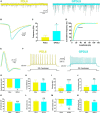
Effects of PDL type on synaptic activity and electrophysiological properties. (A) Example of miniature excitatory post-synaptic currents (mEPSCs) recorded from neurons seeded on PDL6 (20 μg/ml; left yellow trace) and GPDL9 (5 μg/ml; right cyan trace). (B) Average traces of mEPSCs recorded from neurons seeded on PDL6 (20 μg/ml; yellow trace) and GPDL9 (5 μg/ml; cyan trace). Frequency (C) and amplitude (D) of mEPSCs were significantly increased when neurons were seeded on GPDL9 compared to PDL6 (Welch’s test; *p < 0.05; ****p < 0.0001; n = 10–19). (E) Average traces of action potentials elicited near the rheobase in neurons seeded on PDL6 (yellow trace) or GPDL9 (cyan trace). (F) Response of recorded neurons seeded on PDL6 (left yellow trace) or GPDL9 (right cyan trace) to a 1 s depolarizing current pulse (twice the rheobase). Analysis of resting membrane potential [RMP (G)], input resistance [R input (H)], rheobase (I), firing frequency (J), action potential (AP) amplitude (K) and duration (L) and after hyperpolarization (AHP) amplitude (M) and duration (N) in neurons seeded on PDL6 (yellow bars) compared to GPDL9 (cyan bars; t-test; ns p > 0.05; *p < 0.05; **p < 0.01; ***p < 0.001; ****p < 0.0001; n = 10–24).
FIGURE 9
FIGURE 9
Videos 1 and 2: Calcium imaging recorded from cultures seeded on PDL6 (20 μg/ml left) or GPDL9 (5 μg/ml right), infected at DIV2 and imaged at DIV9. Scale bar = 100 μm. Videos are available online as Supplementary Files.
FIGURE 10
FIGURE 10
The PDL substrate influences neuronal network activity. (A) Raw fluorescence of neurons seeded on PDL6 [(a) left upper panel] or GPDL9 [(b) right upper panel], and image of maximum ΔF/F values over time, for neurons plated on PDL6 [(c) left lower panel] or GPDL9 [(d) right lower panel]. (B) Average values of the fluorescence within a region of interest of 11 × 11 pixels for 4 selected neurons seeded on PDL6 (left panel) or GPDL9 (right panel). Scale bar = 100 μm.
FIGURE 11
FIGURE 11
Proper adhesion and maturation of cortical neurons in vitro are highly dependent on the substrate. (1) If seeded on glass without any coating, neurons do not adhere, rapidly reaggregate, and their maturation is compromised. (2) When poly-D-lysine (PDL) concentration is too low or when PDL6 is adsorbed on glass coverslips, neurons can adhere, but slyly tend to cluster a few days after seeding. (3) When PDL coating is optimal and stable, especially with covalent binding, neurons adhere and develop expanded and functional synaptic networks. (4) A too high concentration of PDL is also detrimental because neurons do not attach, float, are small and rounded, and maturation cannot occur.

References

    1. Amendola J., Boumedine N., Sangiardi M., El Far O. (2015). Optimization of neuronal cultures from rat superior cervical ganglia for dual patch recording. Sci. Rep. 5:14455. 10.1038/srep14455 - DOI - PMC - PubMed
    1. Argaw A., Duff G., Zabouri N., Cecyre B., Chaine N., Cherif H., et al. (2011). Concerted action of Cb1 cannabinoid receptor and deleted in colorectal cancer in axon guidance. J. Neurosci. 31 1489–1499. 10.1523/JNEUROSCI.4134-09.2011 - DOI - PMC - PubMed
    1. Bordoni R., Consolandi C., Castiglioni B., Busti E., Bernardi L. R., Battaglia C., et al. (2002). Investigation of the multiple anchors approach in oligonucleotide microarray preparation using linear and stem-loop structured probes. Nucleic Acids Res. 30:E34. 10.1093/nar/30.8.e34 - DOI - PMC - PubMed
    1. Chanda S., Hale W. D., Zhang B., Wernig M., Sudhof T. C. (2017). Unique versus redundant functions of neuroligin genes in shaping excitatory and inhibitory synapse properties. J. Neurosci. 37 6816–6836. 10.1523/JNEUROSCI.0125-17.2017 - DOI - PMC - PubMed
    1. Chen T. W., Wardill T. J., Sun Y., Pulver S. R., Renninger S. L., Baohan A., et al. (2013). Ultrasensitive fluorescent proteins for imaging neuronal activity. Nature 499 295–300. 10.1038/nature12354 - DOI - PMC - PubMed

LinkOut - more resources