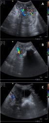
Characterization of uterine involution using B-mode ultrasonography, color Doppler and elastography (acoustic radiation force impulse) for assessing postpartum in Santa Inês ewes
- PMID: 37416868
- PMCID: PMC10321682
- DOI: 10.1590/1984-3143-AR2022-0110
Characterization of uterine involution using B-mode ultrasonography, color Doppler and elastography (acoustic radiation force impulse) for assessing postpartum in Santa Inês ewes
Abstract
The aim of this study was to investigate uterine involution using ultrasonography techniques during postpartum. Postpartum ultrasonography evaluation (B-mode, color Doppler and Acoustic Radiation Force Impulse elastography) of the uterus was performed by transabdominal approach at immediate after birth and sequentially every 48 hours, during 30 days. The uterine echotexture did not present significant variations (P >0.05) being homogeneous in most evaluations; echogenicity of the uterus increased along the evaluation period (P =0.0452). Progressive and remarkable decrease of the total uterine diameter (UD) were observed (P <0.0001), especially during the first days postpartum. The thickness of uterine wall gradually decreased, as well the endometrial, myometrium and lumen diameters (P <0.0001). Uterine blood flow was assessed by Doppler and decreased during postpartum period, being significantly lower (P=0.0225) on the 30th day of postpartum. Uterine parenchyma presented as homogeneous dark areas (not deformable) on qualitative ultrasound elastography and the means shear velocity values of the uterine wall on quantitative elastography did not differ. This is the first study that evaluate the stiffness of uterine wall in healthy ewes, providing baseline data about quantitative and qualitative stiffness of the normal uterus, and it may be a useful tool for early diagnosis of uterine alterations during the postpartum period, using the reference parameter established for the assessment of uterine integrity during postpartum period.
Keywords: puerperium; sheep; uterine involution; uterine stiffness.
Conflict of interest statement
Conflicts of interest: The authors have no conflict of interest to declare.
Figures


Similar articles
-
Assessment of uterine caruncles, uterine cervix, and vulva during the postpartum period in Kivircik ewes with shear-wave elastography.Front Vet Sci. 2024 Dec 31;11:1484189. doi: 10.3389/fvets.2024.1484189. eCollection 2024. Front Vet Sci. 2024. PMID: 39811147 Free PMC article.
-
Quantitative ultrasound elastography and biometry of the bitch uterus in the early puerperium after vaginal delivery and caesarean section.Reprod Domest Anim. 2020 Mar;55(3):364-373. doi: 10.1111/rda.13633. Epub 2020 Jan 31. Reprod Domest Anim. 2020. PMID: 31945229
-
[Continuous, subjective sonographic grayscale analysis of uterine involution in sows during the postpartum period].Tierarztl Prax Ausg G Grosstiere Nutztiere. 2022 Feb;50(1):38-45. doi: 10.1055/a-1741-7855. Epub 2022 Mar 2. Tierarztl Prax Ausg G Grosstiere Nutztiere. 2022. PMID: 35235981 German.
-
The postpartum buffalo: I. Endocrinological changes and uterine involution.Anim Reprod Sci. 2007 Feb;97(3-4):201-15. doi: 10.1016/j.anireprosci.2006.03.004. Epub 2006 Apr 5. Anim Reprod Sci. 2007. PMID: 16600532 Review.
-
Uterine involution: its role in regulating postpartum intervals.J Anim Sci. 1980;51 Suppl 2:16-28. J Anim Sci. 1980. PMID: 6765310 Review.
Cited by
-
Assessment of uterine caruncles, uterine cervix, and vulva during the postpartum period in Kivircik ewes with shear-wave elastography.Front Vet Sci. 2024 Dec 31;11:1484189. doi: 10.3389/fvets.2024.1484189. eCollection 2024. Front Vet Sci. 2024. PMID: 39811147 Free PMC article.
References
-
- Almeida TFAD. Eficiência placentária em ovelhas e consequências para o desenvolvimento e sobrevivência das crias. Uberlândia: Universidade Federal de Uberlândia; 2019. undergraduate thesis. Portuguese.
-
- Badawi ME, Makawi SEA, Abdelghafar RM, Ibrahim MT. Assessment of postpartum uterine involution and progesterone profile in Nubian goats (Capra hircus) J Adv Vet Anim Res. 2014;1(2):36–41. doi: 10.5455/javar.2014.a10. - DOI
LinkOut - more resources
Full Text Sources
