

Pulmonary artery diameter correlates with echocardiographic parameters of right ventricular dysfunction in patients with acute pulmonary embolism
- PMID: 37417082
- PMCID: PMC10401985
- DOI: 10.4103/lungindia.lungindia_357_22
Pulmonary artery diameter correlates with echocardiographic parameters of right ventricular dysfunction in patients with acute pulmonary embolism
Abstract
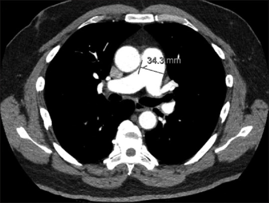
Introduction: Right ventricular dysfunction (RVD) is a key component in the process of risk stratification in patients with acute pulmonary embolism (PE). Echocardiography remains the gold standard for RVD assessment, however, measures of RVD may be seen on CTPA imaging, including increased pulmonary artery diameter (PAD). The aim of our study was to evaluate the association between PAD and echocardiographic parameters of RVD in patients with acute PE.
Methods: Retrospective analysis of patients diagnosed with acute PE was conducted at large academic center with an established pulmonary embolism response team (PERT). Patients with available clinical, imaging, and echocardiographic data were included. PAD was compared to echocardiographic markers of RVD. Statistical analysis was performed using the Student's t test, Chi-square test, or one-way analysis of variance (ANOVA); P < 0.05 was considered statistically significant.
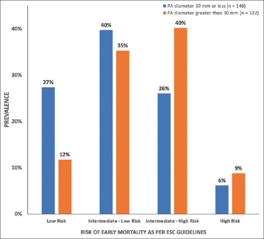
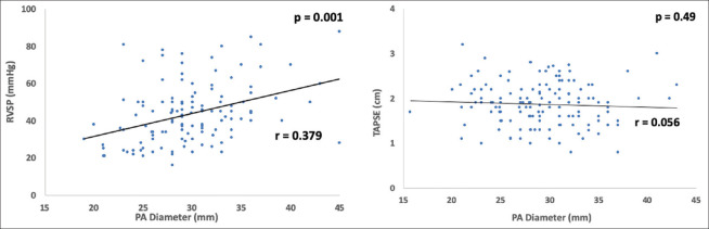
Results: 270 patients with acute PE were identified. Patients with a PAD >30 mm measured on CTPA had higher rates of RV dilation (73.1% vs 48.7%, P < 0.005), RV systolic dysfunction (65.4% vs 43.7%, P < 0.005), and RVSP >30 mmHg (90.2% vs 68%, P = 0.004), but not TAPSE ≤1.6 cm (39.1% vs 26.1%, P = 0.086). A weak increasing linear relationship between PAD and RVSP was noted (r = 0.379, P = 0.001).
Conclusions: Increased PAD in patients with acute PE was significantly associated with echocardiographic markers of RVD. Increased PAD on CTPA in acute PE can serve as a rapid prognostic tool and assist with PE risk stratification at the time of diagnosis, allowing rapid mobilization of a PERT team and appropriate resource utilization.
Keywords: Echocardiography; pulmonary embolism; right ventricular dysfunction.
Conflict of interest statement
There are no conflicts of interest.
Figures
References
-
- Silverstein MD, Heit JA, Mohr DN, Petterson TM, O'Fallon WM, Melton LJ., 3rd Trends in the incidence of deep vein thrombosis and pulmonary embolism:A 25-year population-based study. Arch Intern Med. 1998;158:585–93. - PubMed
-
- Centers for Disease Control and Prevention (CDC) Venous thromboembolism in adult hospitalizations-United States, 2007-2009. MMWR Morb Mortal Wkly Rep. 2012;61:401–4. - PubMed
-
- Konstantinides SV, Meyer G, Becattini C, et al. 2019 ESC Guidelines for the diagnosis and management of acute pulmonary embolism developed in collaboration with the European Respiratory Society (ERS) Eur Heart J. 2020;41:543–603. doi:10.1093/eurheartj/ehz405. - PubMed
LinkOut - more resources
Full Text Sources
