Frontal osteomyelitis post-COVID-19 associated mucormycosis
- PMID: 37417145
- PMCID: PMC10491079
- DOI: 10.4103/IJO.IJO_3117_22
Frontal osteomyelitis post-COVID-19 associated mucormycosis
Abstract
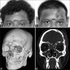
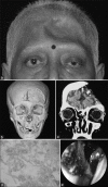
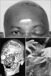
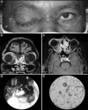
Rhino-orbito-cerebral mucormycosis (ROCM) is the most commonly noted form of mucormycosis, which is the most common secondary fungal infection following severe acute respiratory syndrome coronavirus 2 (SARS-CoV-2) infection. Osteomyelitis is one of the rare sequelae of ROCM, frontal osteomyelitis being the rarest. We present four patients of coronavirus disease 2019 (COVID-19)-associated mucormycosis, who presented with frontal bone osteomyelitis after being treated for ROCM surgically and medically. This is the first case series highlighting this complication in post-COVID-19 mucormycosis patients and needs utmost attention as it can be life-threatening and can cause extreme facial disfiguration. All four patients are alive with salvage of the affected globe and vision being preserved in one patient. If identified early, disfiguration of face and intracranial extension can be avoided.
Keywords: Frontal bone osteomyelitis; Pott's puffy tumor; fungal orbital and sinus infections; mucormycosis; post–COVID-19 mucormycosis.
Conflict of interest statement
None
Figures


Similar articles
-
Pott's puffy tumor: An unusual complication of rhino-orbito-cerebral mucormycosis.World Neurosurg X. 2024 May 4;23:100387. doi: 10.1016/j.wnsx.2024.100387. eCollection 2024 Jul. World Neurosurg X. 2024. PMID: 38746040 Free PMC article.
-
COVID-19-associated rhino-orbital-cerebral mucormycosis: A systematic review, meta-analysis, and meta-regression analysis.Indian J Pharmacol. 2021 Nov-Dec;53(6):499-510. doi: 10.4103/ijp.ijp_839_21. Indian J Pharmacol. 2021. PMID: 34975140 Free PMC article.
-
Is mucormycosis the end? A comprehensive management of orbit in COVID associated rhino-orbital-cerebral mucormycosis: preserving the salvageable.Eur Arch Otorhinolaryngol. 2023 Feb;280(2):819-827. doi: 10.1007/s00405-022-07620-3. Epub 2022 Sep 2. Eur Arch Otorhinolaryngol. 2023. PMID: 36053359 Free PMC article.
-
Rhino-orbital mucormycosis following COVID-19 in previously non-diabetic, immunocompetent patients.Orbit. 2021 Dec;40(6):499-504. doi: 10.1080/01676830.2021.1960382. Epub 2021 Aug 1. Orbit. 2021. PMID: 34338124
-
Rhino-orbito-cerebral mucormycosis and its resurgence during COVID-19 pandemic: A review.Indian J Ophthalmol. 2023 Jan;71(1):39-56. doi: 10.4103/ijo.IJO_1219_22. Indian J Ophthalmol. 2023. PMID: 36588206 Free PMC article. Review.
Cited by
-
Pott's puffy tumor: An unusual complication of rhino-orbito-cerebral mucormycosis.World Neurosurg X. 2024 May 4;23:100387. doi: 10.1016/j.wnsx.2024.100387. eCollection 2024 Jul. World Neurosurg X. 2024. PMID: 38746040 Free PMC article.
References
-
- Sen M, Honavar SG, Bansal R, Sengupta S, Rao R, Kim U, et al. Epidemiology, clinical profile, management, and outcome of COVID-19-associated rhino-orbital-cerebral mucormycosis in 2826 patients in India-Collaborative OPAI-IJO study on mucormycosis in COVID-19 (COSMIC), report 1. Indian J Ophthalmol. 2021;69:1670–92. - PMC - PubMed
-
- Prabhu RM, Patel R. Mucormycosis and entomophthoramycosis: A review of the clinical manifestations, diagnosis and treatment. Clin Microbiol Infect. 2004;10:31–47. - PubMed
Publication types
MeSH terms
LinkOut - more resources
Full Text Sources
Medical
Miscellaneous

