. 2023 Sep;28(3):159.
doi: 10.3892/mmr.2023.13046. Epub 2023 Jul 7.

Saikosaponin A protects against uremic toxin indole‑3 acetic acid‑induced damage to the myocardium

Affiliations

Saikosaponin A protects against uremic toxin indole‑3 acetic acid‑induced damage to the myocardium

Cheng Chen et al. Mol Med Rep. 2023 Sep.

Abstract

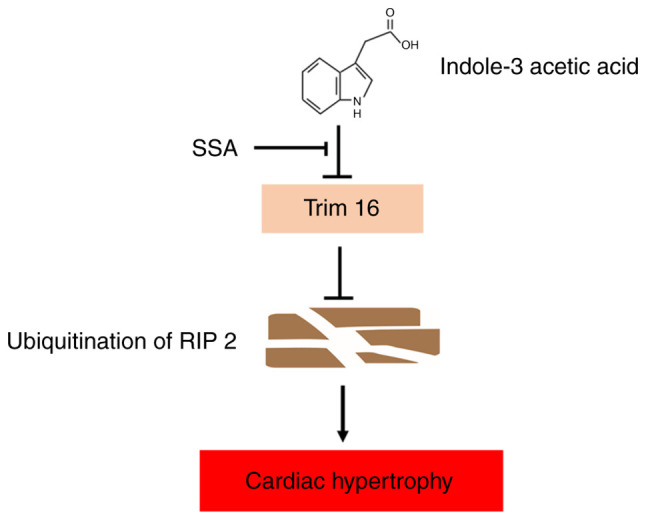
Chronic kidney disease (CKD)‑associated cardiac injury is a common complication in patients with CKD. Indole‑3 acetic acid (IAA) is a uremic toxin that injures the cardiovascular system. Saikosaponin A (SSA) protects against pressure overload‑induced cardiac fibrosis. However, the role and molecular mechanisms of IAA and SSA in CKD‑associated cardiac injury remain unclear. The present study investigated the effects of IAA and SSA on CKD‑associated cardiac injury in neonatal mouse cardiomyocytes and a mouse model of CKD. The expression of tripartite motif‑containing protein 16 (Trim16), receptor interacting protein kinase 2 (RIP2) and phosphorylated‑p38 were assessed using western blotting. The ubiquitination of RIP2 was measured by coimmunoprecipitation, and mouse cardiac structure and function were evaluated using hematoxylin and eosin staining and echocardiography. The results demonstrated that, SSA inhibited IAA‑induced cardiomyocyte hypertrophy, upregulated Trim16 expression, downregulated RIP2 expression and decreased p38 phosphorylation. Furthermore, Trim16 mediated SSA‑induced degradation of RIP2 by ubiquitination. In a mouse model of IAA‑induced CKD‑associated cardiac injury, SSA upregulated the protein expression levels of Trim16 and downregulated those of RIP2. Moreover, SSA alleviated heart hypertrophy and diastolic dysfunction in IAA‑treated mice. Taken together, these results suggest that SSA is a protective agent against IAA‑induced CKD‑associated cardiac injury and that Trim16‑mediated ubiquitination‑related degradation of RIP2 and p38 phosphorylation may contribute to the development of CKD‑associated cardiac injury.

Keywords: cardiac injury; chronic kidney disease; indole‑3 acetic acid; saikosaponin A.


Conflict of interest statement

The authors declare that they have no competing interests.

Figures

Figure 1.
Figure 1.
SSA inhibits IAA-induced cardiomyocyte hypertrophy. (A) Cardiomyocytes were treated with different concentrations of IAA (5, 10 and 50 µmol/l) for 48 h and mRNA expression levels of ANP, BNP and β-MHC were analyzed by qPCR. (B) Cardiomyocytes were pretreated with SSA (1, 10 and 30 µmol/l) for 1 h then exposed to IAA (50 µmol/l) for 48 h and mRNA expression levels of ANP, BNP and β-MHC were analyzed by qPCR. *P<0.01 vs. Cntl, @P<0.01 vs. 50 µmol/l IAA group. SSA, saikosaponin A; IAA, indole-3 acetic acid; ANP, atrial natriuretic peptide; BNP, brain natriuretic peptide; β-MHC, β-myosin heavy chain; Cntl, control; qPCR, quantitative PCR.
Figure 2.
Figure 2.
SSA inhibits Trim16 and RIP2 expression in IAA-treated cardiomyocytes. (A) SiTrim16 or scrambled siRNA (siCntl) were transfected into cardiomyocytes and Trim16 protein expression determined by western blotting. (B) Cardiomyocytes were pretreated with SSA (30 µmol/l) for 1 h and then exposed to IAA (50 µmol/l) for 48 h and Trim16 protein expression measured by Western blotting. (C) SiTrim16 or scrambled siRNA (siCntl) was transfected into cardiomyocytes. Cardiomyocytes were pretreated with 30 µmol/l SSA for 1 h and incubated with IAA (50 µmol/l) for 48 h. RIP2 protein expression was determined by western blotting. *P<0.05 vs. IAA-treated group, &P<0.01 vs. siTrim16 group, #P<0.01 vs. siCntl group, @P<0.01 vs. IAA + SSA-treated group. SSA, saikosaponin A; IAA, indole-3 acetic acid; Trim16, tripartite motif-containing protein 16; RIP2, receptor interacting protein kinase 2.
Figure 3.
Figure 3.
Silencing of Trim16 blocks the inhibitory effect of SSA on cardiomyocyte hypertrophy induced by IAA. Cardiomyocytes were transfected with siTrim16 or scrambled siRNA (siCntl), treated with SSA (30 µmol/l) for 1 h then treated with IAA (50 µmol/l) for 48 h. (A) mRNA expression of ANP, BNP and β-MHC were measured by quantitative PCR. (B) Cell size was observed by immunofluorescence using a troponin antibody (magnification, ×400). (C) Expression of p-p38 and t-p38 was measured by western blotting. *P<0.01 vs. siCntl, @P<0.01 vs. IAA + SSA-treated group. SSA, saikosaponin A; IAA, indole-3 acetic acid; Trim16, tripartite motif-containing protein 16; ANP, atrial natriuretic peptide; BNP, brain natriuretic peptide; β-MHC, β-myosin heavy chain; p, phosphorylated; t, total.
Figure 4.
Figure 4.
SSA alleviates the inhibitory effect of IAA on the K48 ubiquitination of RIP2 in cardiomyocytes. (A) Cardiomyocytes were transfected with siTrim16 or scrambled siRNA (siCntl), treated with SSA (30 µmol/l) for 1 h, then incubated with IAA (50 µmol/l) for 48 h. K48 ubiquitination of RIP2 was measured by immunoprecipitation. (B) Expression of RIP2 was measured by western blotting *P<0.01 vs. IAA-treated group, @P<0.05 vs. IAA + SSA-treated group. SSA, saikosaponin A; IAA, indole-3 acetic acid; RIP2, receptor interacting protein kinase 2; p, phosphorylated; t, total; IP, immunoprecipitation; IB, immunoblotting; si, short interfering RNA.
Figure 5.
Figure 5.
SSA treatment ameliorates IAA-induced cardiac hypertrophy in mice. (A) Serum BUN and Cr levels in the control (n=8), IAA (n=8) and IAA + SSA (n=8) groups of mice were analyzed by urease-glutamate dehydrogenase and enzymatic methods, respectively. (B) The mRNA expression of ANP, BNP and β-MHC in the myocardial tissues of control, IAA- IAA + SSA-treated groups of mice were analyzed by quantitative PCR. (C) Representative micrographs (magnification, 10×) of hematoxylin and eosin-stained transverse sections from hearts of control, IAA- and IAA + SSA-treated mice. *P<0.01 vs. IAA-treated group; #P<0.01 vs. Cntl. SSA, Saikosaponin A; IAA, Indole-3 acetic acid; ANP, Atrial natriuretic peptide; BNP, Brain natriuretic peptide; β-MHC, β-myosin heavy chain; Cntl, control; BUN, blood urea nitrogen; Cr, serum creatinine.
Figure 6.
Figure 6.
SSA treatment ameliorates structural and functional abnormalities of the heart in echocardiography of mice treated with IAA, and also regulates expression of Trim16, RIP2 and p-p38 in the heart. (A) Representative M-mode echocardiograms and the mean LVPWs, LVPWd, LVAWs and LVAWd in the control, IAA- and IAA + SSA-treated groups. (B) Representative M-mode echocardiograms and the mean E/A in the control, IAA- and IAA + SSA-treated groups. (C) Expression of Trim16, RIP2, p-p38 and t-p38 measured by western blotting. *P<0.01, **P<0.05 vs. IAA-treated group. SSA, saikosaponin A; IAA, indole-3 acetic acid; Trim16, tripartite motif-containing protein 16; p, phosphorylated; Cntl, control; RIP2, receptor interacting protein kinase 2; LVPWd, left ventricular end-diastolic posterior wall depth; LVPWs, left ventricular end-systolic posterior wall depth; LVAWd, left ventricular end-diastolic anterior wall depth; LVAWs, left ventricular end-systolic anterior wall depth.
Figure 7.
Figure 7.
Schematic representation of the IAA-induced damage in cardiomyocytes. IAA downregulates the expression of Trim16, which resulted in decreased K48 ubiquitination of RIP2 and increased expression of RIP2. Increased RIP2 expression induces cardiomyocyte hypertrophy and damages diastolic dysfunction of the heart. SSA protects cardiomyocytes from the effects of IAA by upregulating Trim16. SSA, saikosaponin A; IAA, indole-3 acetic acid; Trim16, tripartite motif-containing protein 16; RIP2, receptor interacting protein kinase 2.

Similar articles

Cited by

References

    1. Lv JC, Zhang LX. Prevalence and disease burden of chronic kidney disease. Adv Exp Med Biol. 2019;1165:3–15. doi: 10.1007/978-981-13-8871-2_1. - DOI - PubMed
    1. Webster AC, Nagler EV, Morton RL, Masson P. Chronic kidney disease. Lancet. 2017;389:1238–1252. doi: 10.1016/S0140-6736(16)32064-5. - DOI - PubMed
    1. Matsushita K, Ballew SH, Wang AY, Kalyesubula R, Schaeffner E, Agarwal R. Epidemiology and risk of cardiovascular disease in populations with chronic kidney disease. Nat Rev Nephrol. 2022;18:696–707. doi: 10.1038/s41581-022-00616-6. - DOI - PubMed
    1. Patel N, Yaqoob MM, Aksentijevic D. Cardiac metabolic remodelling in chronic kidney disease. Nat Rev Nephrol. 2022;18:524–537. doi: 10.1038/s41581-022-00576-x. - DOI - PubMed
    1. Chen C, Xie C, Xiong Y, Wu H, Wu L, Zhu J, Xing C, Mao H. Damage of uremic myocardium by p-cresyl sulfate and the ameliorative effect of Klotho by regulating SIRT6 ubiquitination. Toxicol Lett. 2022;367:19–31. doi: 10.1016/j.toxlet.2022.06.006. - DOI - PubMed