

Biological Effects of New Titanium Surface Coatings Based on Ionic Liquids and HMGB1: A Cellular and Molecular Characterization in Lewis Rats
- PMID: 37418317
- PMCID: PMC11292580
- DOI: 10.1021/acsbiomaterials.3c00367
Biological Effects of New Titanium Surface Coatings Based on Ionic Liquids and HMGB1: A Cellular and Molecular Characterization in Lewis Rats
Abstract
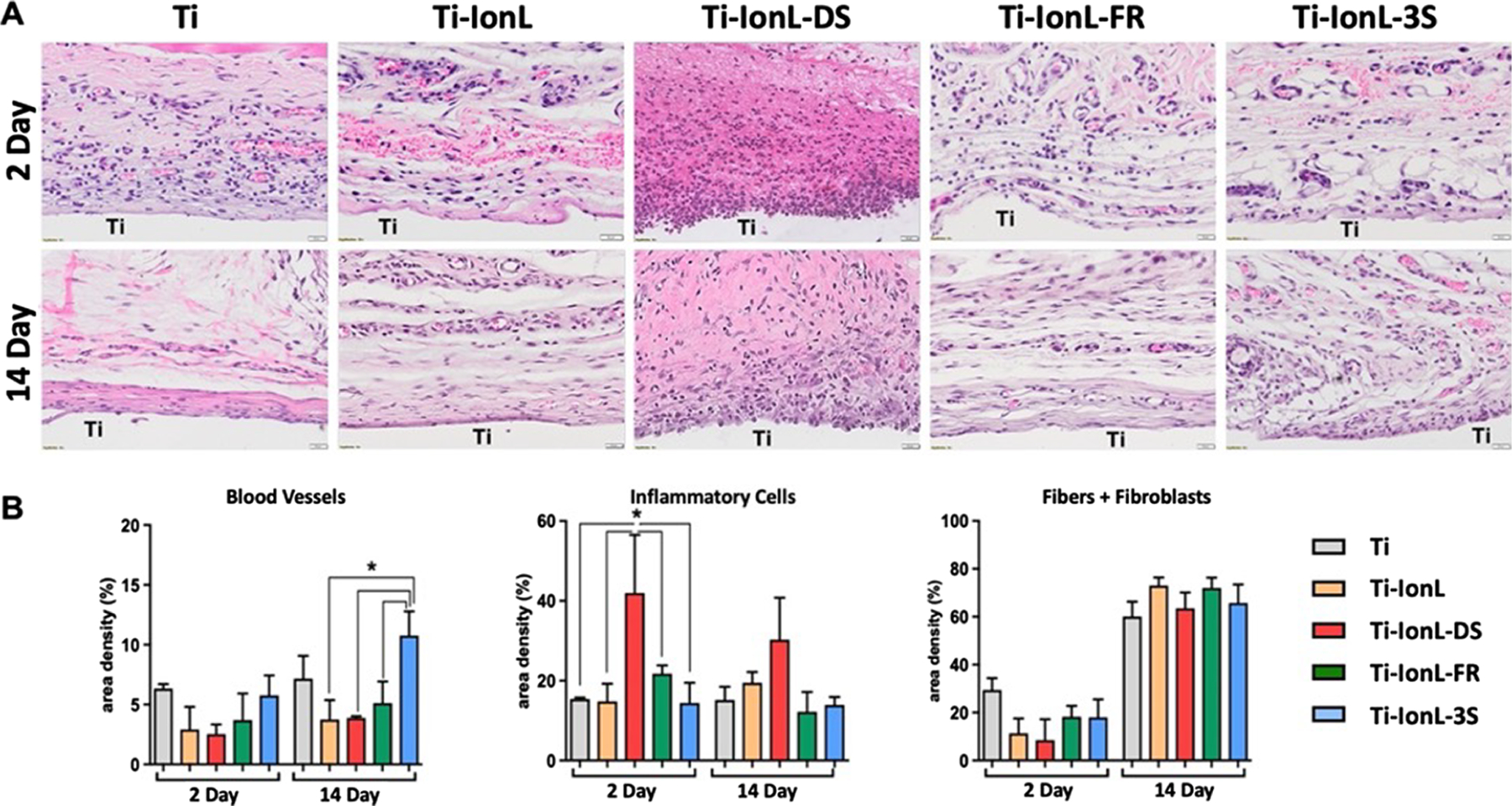
High Mobility Group Box 1 (HMGB1) is a redox-sensitive molecule that plays dual roles in tissue healing and inflammation. We previously demonstrated that HMGB1 is stable when anchored by a well-characterized imidazolium-based ionic liquid (IonL), which serves as a delivery vehicle for exogenous HMGB1 to the site of injury and prevents denaturation from surface adherence. However, HMGB1 exists in different isoforms [fully reduced HMGB1 (FR), a recombinant version of FR resistant to oxidation (3S), disulfide HMGB1 (DS), and inactive sulfonyl HMGB1(SO)] that have distinct biological functions in health and disease. Thus, the goal of this study was to evaluate the effects of different recombinant HMGB1 isoforms on the host response using a rat subcutaneous implantation model. A total of 12 male Lewis rats (12-15 weeks) were implanted with titanium discs containing different treatments (n = 3/time point; Ti, Ti-IonL, Ti-IonL-DS, Ti-IonL-FR, and Ti-IonL-3S) and assessed at 2 and 14 days. Histological (H&E and Goldner trichrome staining), immunohistochemistry, and molecular analyses (qPCR) of surrounding implant tissues were employed for analysis of inflammatory cells, HMGB1 receptors, and healing markers. Ti-IonL-DS samples resulted in the thickest capsule formation, increased pro-inflammatory, and decreased anti-inflammatory cells, while Ti-IonL-3S samples demonstrated suitable tissue healing similar to uncoated Ti discs, as well as an upregulation of anti-inflammatory cells at 14 days compared to all other treatments. Thus, results from this study demonstrated that Ti-IonL-3S are safe alternatives for Ti biomaterials. Future studies are necessary to investigate the healing potential of Ti-IonL-3S in osseointegration scenarios.
Keywords: immunomodulation; implant coating; inflammation; subcutaneous.
Figures


Similar articles
-
Revolutionizing fracture fixation in diabetic and non-diabetic rats: High mobility group box 1-based coating for enhanced osseointegration.Bone. 2023 Dec;177:116917. doi: 10.1016/j.bone.2023.116917. Epub 2023 Sep 20. Bone. 2023. PMID: 37739297 Free PMC article.
-
Effects of Dicationic Imidazolium-Based Ionic Liquid Coatings on Oral Osseointegration of Titanium Implants: A Biocompatibility Study in Multiple Rat Demographics.Genes (Basel). 2022 Apr 2;13(4):642. doi: 10.3390/genes13040642. Genes (Basel). 2022. PMID: 35456448 Free PMC article.
-
Exogenous Protein Delivery of Ionic Liquid-Mediated HMGB1 Coating on Titanium Implants.Langmuir. 2023 Feb 14;39(6):2204-2217. doi: 10.1021/acs.langmuir.2c02688. Epub 2023 Jan 30. Langmuir. 2023. PMID: 36716434
-
Investigation of the early healing response to dicationic imidazolium-based ionic liquids: a biocompatible coating for titanium implants.ACS Biomater Sci Eng. 2020 Feb 10;6(2):984-994. doi: 10.1021/acsbiomaterials.9b01884. Epub 2020 Jan 14. ACS Biomater Sci Eng. 2020. PMID: 32656316 Free PMC article.
-
High Mobility Group Box 1 Expression in Oral Inflammation and Regeneration.Front Immunol. 2020 Jul 14;11:1461. doi: 10.3389/fimmu.2020.01461. eCollection 2020. Front Immunol. 2020. PMID: 32760399 Free PMC article. Review.
Cited by
-
Revolutionizing fracture fixation in diabetic and non-diabetic rats: High mobility group box 1-based coating for enhanced osseointegration.Bone. 2023 Dec;177:116917. doi: 10.1016/j.bone.2023.116917. Epub 2023 Sep 20. Bone. 2023. PMID: 37739297 Free PMC article.
References
-
- Weinstein Allan; Horowitz Emanuel; Ruff Arthur W. Retrieval and Analysis of Orthopaedic Implants. DOI: 10.6028/NBS.SP.472. - DOI
Publication types
MeSH terms
Substances
Grants and funding
LinkOut - more resources
Full Text Sources