

Generation and characterization of two immortalized dermal fibroblast cell lines from the spiny mouse (Acomys)
- PMID: 37418364
- PMCID: PMC10328323
- DOI: 10.1371/journal.pone.0280169
Generation and characterization of two immortalized dermal fibroblast cell lines from the spiny mouse (Acomys)
Abstract
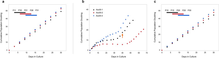
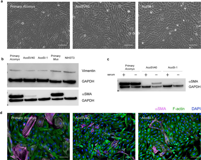
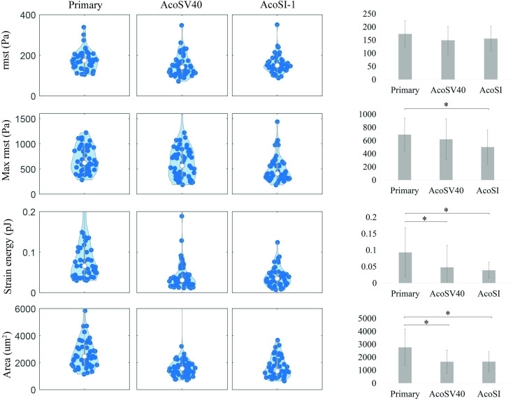
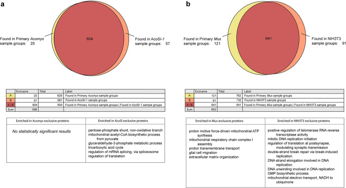
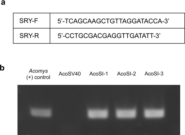
The spiny mouse (Acomys) is gaining popularity as a research organism due to its phenomenal regenerative capabilities. Acomys recovers from injuries to several organs without fibrosis. For example, Acomys heals full thickness skin injuries with rapid re-epithelialization of the wound and regeneration of hair follicles, sebaceous glands, erector pili muscles, adipocytes, and dermis without scarring. Understanding mechanisms of Acomys regeneration may uncover potential therapeutics for wound healing in humans. However, access to Acomys colonies is limited and primary fibroblasts can only be maintained in culture for a limited time. To address these obstacles, we generated immortalized Acomys dermal fibroblast cell lines using two methods: transfection with the SV40 large T antigen and spontaneous immortalization. The two cell lines (AcoSV40 and AcoSI-1) maintained the morphological and functional characteristics of primary Acomys fibroblasts, including maintenance of key fibroblast markers and ECM deposition. The availability of these cells will lower the barrier to working with Acomys as a model research organism, increasing the pace at which new discoveries to promote regeneration in humans can be made.
Copyright: © 2023 Dill et al. This is an open access article distributed under the terms of the Creative Commons Attribution License, which permits unrestricted use, distribution, and reproduction in any medium, provided the original author and source are credited.
Conflict of interest statement
The authors have declared that no competing interests exist.
Figures
Similar articles
-
Optimal skin regeneration after full thickness thermal burn injury in the spiny mouse, Acomys cahirinus.Burns. 2018 Sep;44(6):1509-1520. doi: 10.1016/j.burns.2018.05.018. Epub 2018 Jun 11. Burns. 2018. PMID: 29903601
-
Insights into the regeneration of skin from Acomys, the spiny mouse.Exp Dermatol. 2019 Apr;28(4):436-441. doi: 10.1111/exd.13847. Epub 2019 Jan 15. Exp Dermatol. 2019. PMID: 30457673 Review.
-
Unique behavior of dermal cells from regenerative mammal, the African Spiny Mouse, in response to substrate stiffness.J Biomech. 2018 Nov 16;81:149-154. doi: 10.1016/j.jbiomech.2018.10.005. Epub 2018 Oct 14. J Biomech. 2018. PMID: 30361050
-
Cellular events during scar-free skin regeneration in the spiny mouse, Acomys.Wound Repair Regen. 2016 Jan-Feb;24(1):75-88. doi: 10.1111/wrr.12385. Epub 2016 Jan 19. Wound Repair Regen. 2016. PMID: 26606280
-
Regeneration in the spiny mouse, Acomys, a new mammalian model.Curr Opin Genet Dev. 2020 Oct;64:31-36. doi: 10.1016/j.gde.2020.05.019. Epub 2020 Jun 26. Curr Opin Genet Dev. 2020. PMID: 32599302 Free PMC article. Review.
Cited by
-
Spiny mice (Acomys) have evolved cellular features to support regenerative healing.Ann N Y Acad Sci. 2025 Feb;1544(1):5-26. doi: 10.1111/nyas.15281. Epub 2025 Jan 13. Ann N Y Acad Sci. 2025. PMID: 39805008 Review.
-
Mycobacterium avium inhibits protein kinase C and MARCKS phosphorylation in human cystic fibrosis and non-cystic fibrosis cells.PLoS One. 2024 Oct 16;19(10):e0308299. doi: 10.1371/journal.pone.0308299. eCollection 2024. PLoS One. 2024. PMID: 39413095 Free PMC article.
-
Osteogenic shift in the adipose-derived stem cells of Acomys cahirinus is linked to impaired adipose tissue self-renewal.Front Cell Dev Biol. 2025 Jul 30;13:1603405. doi: 10.3389/fcell.2025.1603405. eCollection 2025. Front Cell Dev Biol. 2025. PMID: 40809694 Free PMC article.
-
Type III Collagen Regulates Matrix Architecture and Mechanosensing during Wound Healing.J Invest Dermatol. 2025 Apr;145(4):919-938.e14. doi: 10.1016/j.jid.2024.08.013. Epub 2024 Sep 3. J Invest Dermatol. 2025. PMID: 39236902
References
-
- Gaire J, Varholick JA, Rana S, Sunshine MD, Doré S, Barbazuk WB, et al.. Spiny mouse (Acomys): an emerging research organism for regenerative medicine with applications beyond the skin. npj Regenerative Medicine 2021. 6:1 [Internet]. 2021 Jan 4 [cited 2021 Jul 20];6(1):1–6. Available from: https://www.nature.com/articles/s41536-020-00111-1. doi: 10.1038/s41536-020-00111-1 - DOI - PMC - PubMed
-
- Seifert AW, Kiama SG, Seifert MG, Goheen JR, Palmer TM, Maden M. Skin shedding and tissue regeneration in African spiny mice (Acomys). Nature 2012. 489:7417 [Internet]. 2012 Sep 26 [cited 2022 Jan 31];489(7417):561–5. Available from: https://www.nature.com/articles/nature11499. doi: 10.1038/nature11499 - DOI - PMC - PubMed
-
- Brant JO, Lopez MC, Baker H v., Barbazuk WB, Maden M. A Comparative Analysis of Gene Expression Profiles during Skin Regeneration in Mus and Acomys. PLoS One [Internet]. 2015. Nov 1 [cited 2022 Jan 31];10(11):e0142931. Available from: https://journals.plos.org/plosone/article?id=10.1371/journal.pone.0142931. doi: 10.1371/journal.pone.0142931 - DOI - PMC - PubMed
-
- Brant JO, Yoon JH, Polvadore T, Barbazuk WB, Maden M. Cellular events during scar-free skin regeneration in the spiny mouse, Acomys. Wound Repair and Regeneration [Internet]. 2016. Jan 1 [cited 2022 Jan 31];24(1):75–88. Available from: https://onlinelibrary.wiley.com/doi/full/10.1111/wrr.12385. - DOI - PubMed
Publication types
MeSH terms
Grants and funding
LinkOut - more resources
Full Text Sources
Research Materials
