. 2023 Jul 7;18(7):e0280070.
doi: 10.1371/journal.pone.0280070. eCollection 2023.

Bacterial cytological profiling reveals interactions between jumbo phage φKZ infection and cell wall active antibiotics in Pseudomonas aeruginosa

Affiliations

Bacterial cytological profiling reveals interactions between jumbo phage φKZ infection and cell wall active antibiotics in Pseudomonas aeruginosa

Hannah Tsunemoto et al. PLoS One. .

Abstract

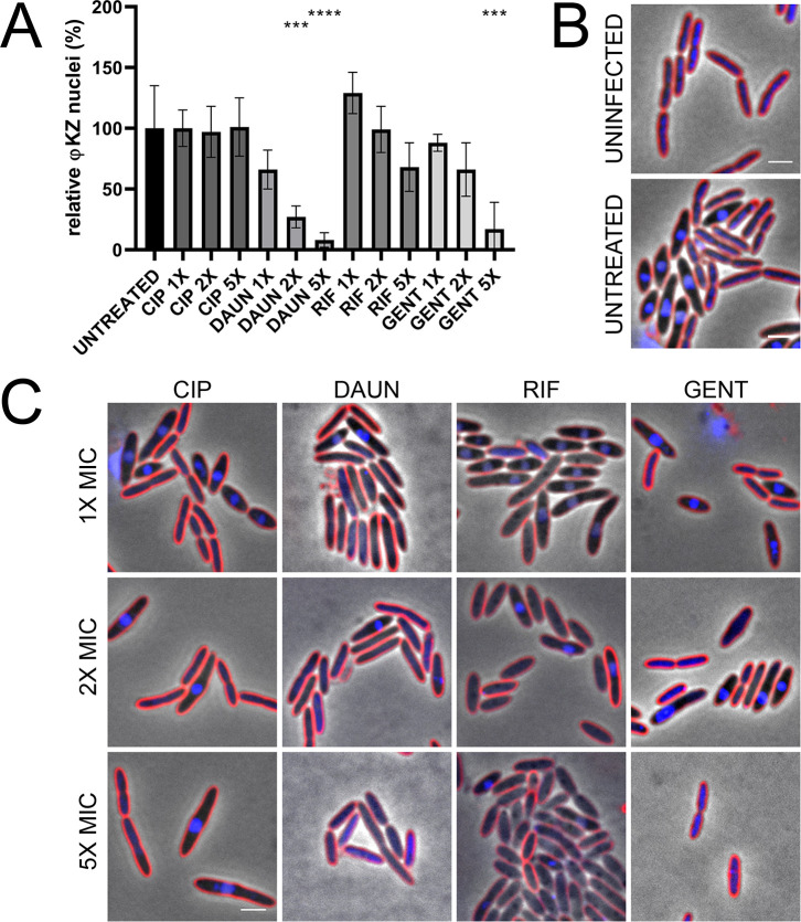
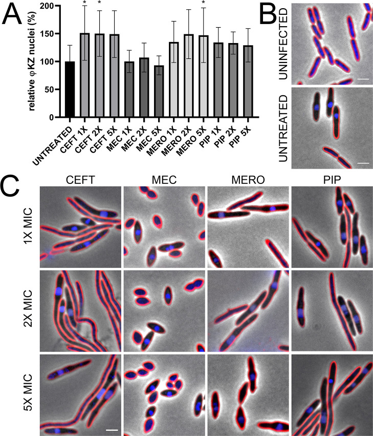
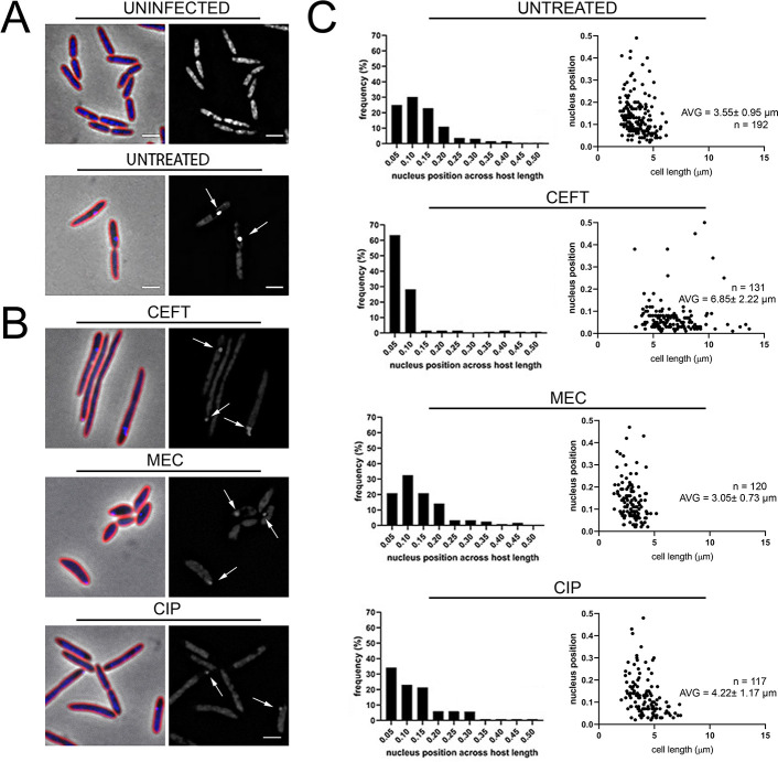
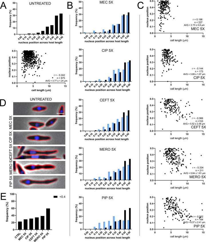
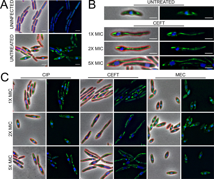
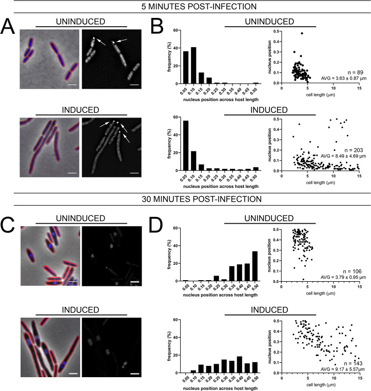
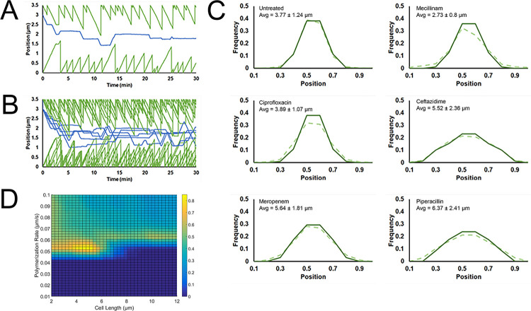
The emergence of antibiotic resistance in bacteria has led to the investigation of alternative treatments, such as phage therapy. In this study, we examined the interactions between the nucleus-forming jumbo phage ФKZ and antibiotic treatment against Pseudomonas aeruginosa. Using the fluorescence microscopy technique of bacterial cytological profiling, we identified mechanism-of-action-specific interactions between antibiotics that target different biosynthetic pathways and ФKZ infection. We found that certain classes of antibiotics strongly inhibited phage replication, while others had no effect or only mildly affected progression through the lytic cycle. Antibiotics that caused an increase in host cell length, such as the cell wall active antibiotic ceftazidime, prevented proper centering of the ФKZ nucleus via the PhuZ spindle at midcell, leading us to hypothesize that the kinetic parameters of the PhuZ spindle evolved to match the average length of the host cell. To test this, we developed a computational model explaining how the dynamic properties of the PhuZ spindle contribute to phage nucleus centering and why some antibiotics affect nucleus positioning while others do not. These findings provide an understanding of the molecular mechanisms underlying the interactions between antibiotics and jumbo phage replication.


Conflict of interest statement

The authors have declared that no competing interests exist.

Figures

Fig 1
Fig 1. Treatment of P. aeruginosa K2733 with different antibiotics show differential φKZ infection related to MOA and host cell phenotype at 30 min post-infection.
(A) Quantification of phage infection (presence of distinct phage nuclei) under treatment conditions, relative to the untreated infected control. Error bars represent standard deviation of biological triplicates. *** = p < 0.0005, **** = p < 0.00005 (B) Microscopy of uninfected and untreated infected controls, and (C) treated infected samples: ciprofloxacin (CIP), daunorubicin (DAUN), rifampicin (RIF), and gentamicin (GENT). Cell membrane strained with FM4-64 (red) and DNA stained with DAPI (blue). Scale bar represents 2 μm.
Fig 2
Fig 2. Treatment of P. aeruginosa K2733 with cell wall active antibiotics show differential φKZ infection related to MOA and host cell phenotype at 30 min post-infection.
(A) Quantification of phage infection (presence of distinct phage nuclei) under treatment conditions, relative to the untreated infected control. Error bars represent standard deviation of biological triplicates. * = p < 0.05 (B) Microscopy of uninfected and untreated infected controls, and (C) treated infected samples: ceftazidime (CEFT), mecillinam (MEC), meropenem (MERO), and piperacillin (PIP). Cell membrane strained with FM4-64 (red) and DNA stained with DAPI (blue). Scale bar represents 2 μm.
Fig 3
Fig 3. Treatment with CEFT increases early φKZ binding and infection.
(A) Microscopy of uninfected and untreated infected controls, and (B) treated infected samples with 5X MIC at 5 min post-infection. Cell membrane strained with FM4-64 (red) and DNA stained with DAPI (blue). Scale bar represents 2 μm. White arrows indicate phage DNA at early stages of infection. (C) Nucleus position across P. aeruginosa host length of untreated and treated (5X MIC) infected samples. Histograms show early phage nucleus position across normalized host cell length. Scatterplots show early phage nucleus position across relative host cell length. Average cell length for each treatment condition shown.
Fig 4
Fig 4. Histogram of nucleus position across normalized P.
aeruginosa host cell length for (A) untreated infected control and for (B) treated samples at 5X MIC, in blue, at 30 min post-infection. Black bars represent untreated infected control and are included for comparison. Scatterplots show nucleus position for across relative P. aeruginosa host cell length for (A) untreated infected control and for (C) treated samples at 5X MIC, along with average cell length for each treatment condition. Correlation coefficients between average cell length and nucleus position (r) shown for each treatment condition. (D) Representative cell images of untreated infected controls and treated infected samples. Cell membrane strained with FM4-64 (red) and DNA stained with DAPI (blue). Scale bar represents 2 μm. (E) Frequency of nucleus position out of the middle 20% of the host cell for untreated and treated samples at 5X MIC.
Fig 5
Fig 5. Antibiotic treatment leads to aberrant PhuZ filament and spindle dynamics at 30 min post-infection.
(A) Microscopy of uninfected and untreated infected controls. (B) Representative cell images of untreated infected controls, and CEFT treated infected samples at all concentrations. (C) Microscopy of treated infected samples. Cell membrane strained with FM4-64 (red) and DNA stained with DAPI (blue). GFP-PhuZ (green) under 0.1% arabinose induction. Scale bar represents 2 μm.
Fig 6
Fig 6. Over-expression of the cell division inhibitor protein sulA leads to increased cell length but not increased φKZ infection rates.
(A,C) Microscopy of infected uninduced or induced sulA cells at (A) 5 min or (C) 30 min post-infection. Cell membrane strained with FM4-64 (red) and DNA stained with DAPI (blue). Scale bar represents 2 μm. White arrows indicate phage DNA at early stages of infection. (B,D) Histogram of phage nucleus position across normalized host cell length at (B) 5 min or (D) 30 min post-infection. Scatterplots (D) show nucleus position for across relative P. aeruginosa host cell length at 30 min post-infection for uninduced or induced sulA cells, along with average cell length for induced and uninduced conditions.
Fig 7
Fig 7. Computational modeling of PhuZ dynamics under antibiotic treatment.
(A) A single run of the model showing filament end positions in green and the position of the phage nucleus center in blue. The cell length was set at 3.5 μm and the simulation ran for 30 minutes. (B) Five overlapping traces of the model using parameters identical to (A) provide a visualization of the stochastic behavior of the model. (C) Histograms of phage nucleus position after 30 minutes has elapsed for both the model (solid line) and the measured data (dotted line). All model parameters were kept the same with the exception of cell length which was sampled from a normal distribution with mean and standard deviation given by each treatment condition. (D) Heatmap indicating combinations of cell length and polymerization rate that lead to centering of the phage nucleus. Yellow indicates that a high (>80%) percentage of the phage nuclei positions were located within the central 20% of the cell while blue indicates a low percentage. The effect of all parameters reveals a narrow window in which the phage nucleus can be effectively centered.

References

    1. Thomas J.A., Rolando M.R., Carroll C.A., Shen P.S., Belnap D.M., Weintraub S.T., et al.., Characterization of Pseudomonas chlororaphis myovirus 201varphi2-1 via genomic sequencing, mass spectrometry, and electron microscopy. Virology, 2008. 376(2): p. 330–8. doi: 10.1016/j.virol.2008.04.004 - DOI - PMC - PubMed
    1. Monson R., Foulds I., Foweraker J., Welch M., and Salmond G.P.C., The Pseudomonas aeruginosa generalized transducing phage phiPA3 is a new member of the phiKZ-like group of ’jumbo’ phages, and infects model laboratory strains and clinical isolates from cystic fibrosis patients. Microbiology (Reading), 2011. 157(Pt 3): p. 859–867. doi: 10.1099/mic.0.044701-0 - DOI - PubMed
    1. Krylov V.N., Dela Cruz D.M., Hertveldt K., and Ackermann H.W., “φKZ-like viruses”, a proposed new genus of myovirus bacteriophages. Archives of Virology, 2007. 152(10): p. 1955–1959. - PubMed
    1. Mesyanzhinov V.V., Robben J., Grymonprez B., Kostyuchenko V.A., Bourkaltseva M.V., Sykilinda N.N., et al.., The genome of bacteriophage phiKZ of Pseudomonas aeruginosa. J Mol Biol, 2002. 317(1): p. 1–19. doi: 10.1006/jmbi.2001.5396 - DOI - PubMed
    1. Chaikeeratisak V., Khanna K., Nguyen K.T., Sugie J., Egan M.E., Erb M.L., et al.., Viral Capsid Trafficking along Treadmilling Tubulin Filaments in Bacteria. Cell, 2019. 177(7): p. 1771–1780 e12. doi: 10.1016/j.cell.2019.05.032 - DOI - PMC - PubMed

Publication types

MeSH terms