. 2023 Jul 7;18(7):e0288162.
doi: 10.1371/journal.pone.0288162. eCollection 2023.

Pathogen and human NDPK-proteins promote AML cell survival via monocyte NLRP3-inflammasome activation

Affiliations

Pathogen and human NDPK-proteins promote AML cell survival via monocyte NLRP3-inflammasome activation

Sandro Trova et al. PLoS One. .

Abstract

A history of infection has been linked with increased risk of acute myeloid leukaemia (AML) and related myelodysplastic syndromes (MDS). Furthermore, AML and MDS patients suffer frequent infections because of disease-related impaired immunity. However, the role of infections in the development and progression of AML and MDS remains poorly understood. We and others previously demonstrated that the human nucleoside diphosphate kinase (NDPK) NM23-H1 protein promotes AML blast cell survival by inducing secretion of IL-1β from accessory cells. NDPKs are an evolutionary highly conserved protein family and pathogenic bacteria secrete NDPKs that regulate virulence and host-pathogen interactions. Here, we demonstrate the presence of IgM antibodies against a broad range of pathogen NDPKs and more selective IgG antibody activity against pathogen NDPKs in the blood of AML patients and normal donors, demonstrating that in vivo exposure to NDPKs likely occurs. We also show that pathogen derived NDPK-proteins faithfully mimic the catalytically independent pro-survival activity of NM23-H1 against primary AML cells. Flow cytometry identified that pathogen and human NDPKs selectively bind to monocytes in peripheral blood. We therefore used vitamin D3 differentiated monocytes from wild type and genetically modified THP1 cells as a model to demonstrate that NDPK-mediated IL-1β secretion by monocytes is NLRP3-inflammasome and caspase 1 dependent, but independent of TLR4 signaling. Monocyte stimulation by NDPKs also resulted in activation of NF-κB and IRF pathways but did not include the formation of pyroptosomes or result in pyroptotic cell death which are pivotal features of canonical NLRP3 inflammasome activation. In the context of the growing importance of the NLRP3 inflammasome and IL-1β in AML and MDS, our findings now implicate pathogen NDPKs in the pathogenesis of these diseases.


Conflict of interest statement

The authors have declared that no competing interests exist.

Figures
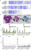
Fig 1
Fig 1. NM23-H1 and pathogen NDPKs are highly conserved and recognized by human humoral antibody responses.
(A) NDPK protein sequences alignments were obtained with ClustalW and visualised with JalView by Percentage Identity. Red * indicates the key residues for NM23-H1 enzymatic activities and structural organization. (B) 3D alignment of NM23-H1 (blue) crystal structure (pdb 1JXV) to S.aureus NDPK (red) tetramer and monomer (pdb 3Q83) and E.coli (green) monomer (pdb 2HUR) were generated with PyMol 2.3 (See also S1 Table). (C) Representative data of four independent samples (#86 and 44 for IgM; #65 and 12 for IgG), two with high CRP (red bars) and two with low CRP (green bars). Averages are expressed as mean ± SEM. (D) Dot plots for all the analyzed samples; black dots: AML samples; red dots: Normal donor samples. Grey bar indicates median ± interquartiles. All samples measured for IgM activity were also measured for IgG activity. CRP values and individual antibody activities for all samples are summarized in S2 Table.
Fig 2
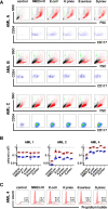
Fig 2. Pathogen NDPKs mimic the action of NM23-H1 against primary AML cells.
(A) Representative flow cytometer plots for three primary AML cells treated with rNDPKs for 7 days. Live (in red) and dead cells (in black) were identified by Forward Scatter (FSC) and Side Scatter (SSC). Counting beads (in green) were used to enumerate live cells. Immunostaining with CD34 and CD117 identified the AML blasts amongst live cells. (B) Dot plots show the mean ± SEM of at least 3 replicates for live cells/ml (red circles) and live blasts/ml (blue squares). Dotted line is number of total viable cells at day 0. (C) Flow cytometry histograms of AML 4 cells stained with propidium iodide for cell cycle analysis. Bars and percentages represent cells in S/G2M phase. AML 2 and AML 4 treatments are all statistically different to the controls; p<0.05, One-way ANOVA with Dunnet’s test. Gating strategy is presented in S1 Fig.
Fig 3
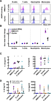
Fig 3. Promotion of AML cell survival by both human and pathogen rNDPKs is mediated via interaction with non-malignant myeloid cells.
(A) Whole blood from normal donors was diluted and incubated with fluorescently labelled BSA control (in blue), rNM23-H1 (in pink) and S.pneumoniae rNDPKR (in grey). Representative plots for B-cells (CD19), T-cells (CD3), neutrophils (CD11b+CD14-) and monocytes (CD11b+CD14+) are shown (See S2 Fig for gating strategy). (B) Fluorescence geometric mean fold changes compared to BSA control of each individual cell type for n = 4 normal donors. (C) ELISA for IL-1β and IL-6 cytokines in conditioned media (CM) generated incubating for 18 hours diluted whole blood (closed circles) or PBMC (open squares) with rNM23-H1 or rNDPK from S.pneumoniae and C.albicans. (D) Red cells from normal donor’s whole blood were lysed and leukocytes were depleted from CD14+ cells (monocytes). Either total white cells or monocyte depleted cells were treated with NM23-H1 or rNDPK for 18h and IL-1β and IL-6 were analyzed by ELISA.
Fig 4
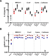
Fig 4. Induction of cytokines and preservation of AML cell viability does not require enzymatic activity of human and pathogen NDPKs.
(A) Vitamin D3 differentiated THP-1 were incubated for 18 hours in presence of WT (in red) and mutated rNDPK (in black), conditioned media was harvested and IL-1β was measured by ELISA. (B) AML10 was treated for 7 days with WT or mutated rNDPK and cell viability of total cells and blasts was measured by flow cytometry. Dot plots show the mean ± SEM of at least 3 replicates for live cells/ml (red circles) and live blasts/ml (blue squares). All the WT and mutants rNDPK treatments are statistically different from the control; p<0.05, One-way ANOVA with Dunnet’s test. Averages are expressed as mean ± SEM.
Fig 5
Fig 5. NM23-H1 and pathogen NDPKs activate the NLRP3 inflammasome pathway.
(A) Whole blood from normal donors was diluted 2:3 and incubated with nigericin, LPS and rNDPKs. The caspase-1 fluorescent dye FLICA 660 was added after 60’ and incubation continued for further 90’. Red cells were lysed, and leukocytes were immunophenotyped by flow cytometry. * p<0.05 to controls, One-way ANOVA with Dunnet’s test. (B) Caspase-1 specific activity measured with the Caspase-Glo 1 Bioluminescent Inflammasome Assay for both Vitamin D3 differentiated THP-1 wild type (WT, blue circles) and Caspase-1 deficient (CASP1def, pink squares) treated for 150’with nigericin, LPS and rNDPKs. Luminescence of the Ac-YVAD-CHO inhibited reactions was subtracted from the total luminescence. * p<0.05 to controls within same cell line, # p<0.05 between WT and CASP1def; One-way ANOVA with post-hoc Tukey’s test. (C) IL-1β ELISA of the conditioned media of Vitamin D3 differentiated THP-1 wild type (WT, in blue), in presence or absence of the Caspase-1 inhibitor Ac-YVAD-cmk (light blue), and of the THP-1 Caspase-1 deficient cell line (pink), treated for 18h with nigericin, LPS or rNDPKs. (D) IL-1β ELISA of the conditioned media from Vitamin D3 differentiated THP-1 wild type (WT, in blue), THP-1 NLRP3 deficient (NLRP3def, in green) and THP-1 NLRP3 knockout (in red), in presence or absence of the NLRP3 inhibitor MCC950 (light blue and green), treated for 18h with nigericin, LPS or rNDPKs. Averages are expressed as mean ± SEM. * p<0.05 to controls, One-way ANOVA with Dunnet’s test.
Fig 6
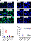
Fig 6. NM23-H1 and pathogen NDPKs activation of the NLRP3 inflammasome pathway occurs without pyroptosome formation.
(A) Vitamin D3 differentiated THP-1 ASC::GFP cells were treated for 6 hours with LPS and rNDPKs or for 3 hours with LPS to induce ASC::GFP protein with subsequent treatment with Nigericin for further 3 hours (LPS+Nig). ASC::GFP protein was visualized by immunofluorescent microscopy. (B) Immunofluorescent staining of ASC specks in Vitamin D3 differentiated wtTHP-1. Arrows indicate ASC specks. (C) Dot plot of the quantification of the number of ASC specks per total nuclei in THP-1 wild type. * p<0.05 compared to control, One-way ANOVA with Dunnet’s test within the same cell line. (D) Dot plot of the viability of Vitamin D3 differentiated THP-1 WT (WT, in blue), NLRP3 deficient (NLRP3def, in green) and Caspase-1 deficient (CASP1def, in pink) after treatment with nigericin, LPS, and rNDPKs. Viability was measured by flow cytometry and the Forward Scatter/Side Scatter parameters. * p<0.05, One-way ANOVA with Dunnet’s test within the same cell line. Averages are expressed as mean ± SEM.
Fig 7
Fig 7. NDPK induced Activation of NF-κB, secretion of IL-1β and IRF activation occur independently of TLR4 signaling.
THP1-Dual (black closed circles) and THP1-Dual TLR4 KO (red open circles) were differentiated into monocytes with Vitamin D3. Following 18 hours exposure to the treatments shown, the following were measured: (A) NF-κB-SEAP reporter gene activity. (B) IL-1β ELISA of the conditioned media, and (C) IRF-Lucia-Luciferase reporter gene activity. (D) Data for IRF Lucia-Luciferase reporter gene activity were plotted against IL-1β ELISA results for Vitamin D3-differentiated THP1-Dual (black) and THP1-Dual TLR4 KO (red) monocytes. Line of fit is log10. PMB; polymyxin-B. TAK; TAK-242. Data are the mean of n = 4–6 ± SEM.

References

    1. Kristinsson SY, Bjorkholm M, Hultcrantz M, Derolf AR, Landgren O, Goldin LR. Chronic immune stimulation might act as a trigger for the development of acute myeloid leukemia or myelodysplastic syndromes. J Clin Oncol. 2011;29(21):2897–903. Epub 2011/06/22. doi: 10.1200/JCO.2011.34.8540 ; PubMed Central PMCID: PMC3138717. - DOI - PMC - PubMed
    1. Hamalainen S, Kuittinen T, Matinlauri I, Nousiainen T, Koivula I, Jantunen E. Neutropenic fever and severe sepsis in adult acute myeloid leukemia (AML) patients receiving intensive chemotherapy: Causes and consequences. Leuk Lymphoma. 2008;49(3):495–501. Epub 2008/02/26. doi: 10.1080/10428190701809172 . - DOI - PubMed
    1. Dayyani F, Conley AP, Strom SS, Stevenson W, Cortes JE, Borthakur G, et al.. Cause of death in patients with lower-risk myelodysplastic syndrome. Cancer. 2010;116(9):2174–9. Epub 2010/02/18. doi: 10.1002/cncr.24984 ; PubMed Central PMCID: PMC3753205. - DOI - PMC - PubMed
    1. Nachtkamp K, Stark R, Strupp C, Kundgen A, Giagounidis A, Aul C, et al.. Causes of death in 2877 patients with myelodysplastic syndromes. Ann Hematol. 2016;95(6):937–44. Epub 2016/03/31. doi: 10.1007/s00277-016-2649-3 . - DOI - PubMed
    1. Halpern AB, Culakova E, Walter RB, Lyman GH. Association of Risk Factors, Mortality, and Care Costs of Adults With Acute Myeloid Leukemia With Admission to the Intensive Care Unit. JAMA Oncol. 2017;3(3):374–81. Epub 2016/11/11. doi: 10.1001/jamaoncol.2016.4858 ; PubMed Central PMCID: PMC5344736. - DOI - PMC - PubMed

MeSH terms

Substances