. 2023 Jul 7;9(27):eadi0263.
doi: 10.1126/sciadv.adi0263. Epub 2023 Jul 7.

Multiplex enzymatic synthesis of DNA with single-base resolution

Affiliations

Multiplex enzymatic synthesis of DNA with single-base resolution

Damiano Verardo et al. Sci Adv. .

Abstract

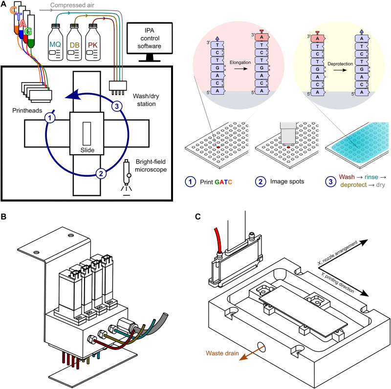
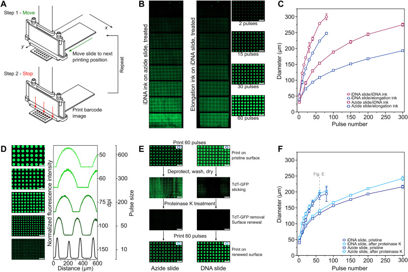
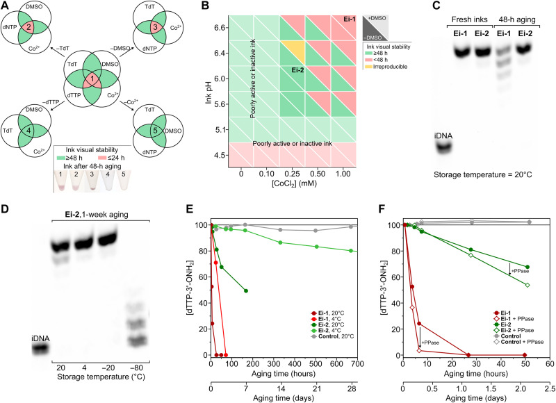
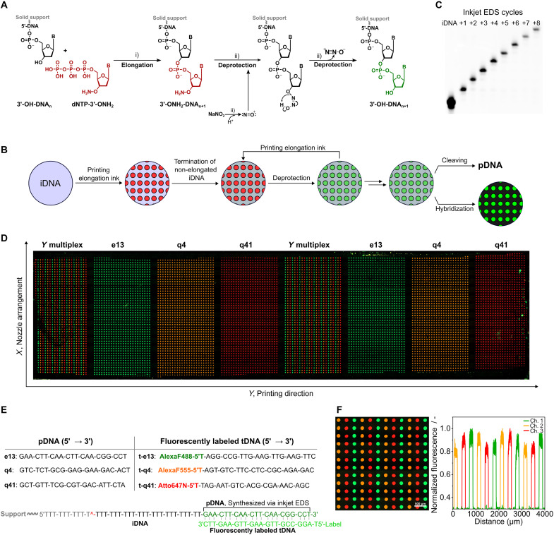
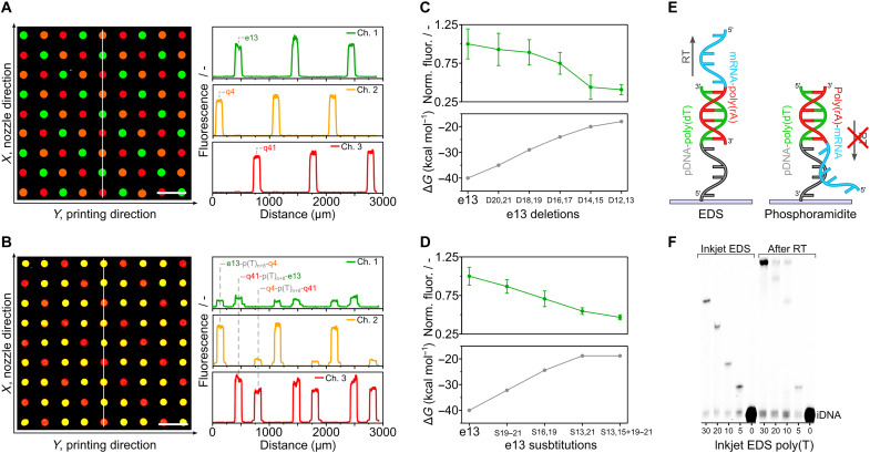
Enzymatic DNA synthesis (EDS) is a promising benchtop and user-friendly method of nucleic acid synthesis that, instead of solvents and phosphoramidites, uses mild aqueous conditions and enzymes. For applications such as protein engineering and spatial transcriptomics that require either oligo pools or arrays with high sequence diversity, the EDS method needs to be adapted and certain steps in the synthesis process spatially decoupled. Here, we have used a synthesis cycle comprising a first step of site-specific silicon microelectromechanical system inkjet dispensing of terminal deoxynucleotidyl transferase enzyme and 3' blocked nucleotide, and a second step of bulk slide washing to remove the 3' blocking group. By repeating the cycle on a substrate with an immobilized DNA primer, we show that microscale spatial control of nucleic acid sequence and length is possible, which, here, are assayed by hybridization and gel electrophoresis. This work is distinctive for enzymatically synthesizing DNA in a highly parallel manner with single base control.


Figures

Fig. 1.
Fig. 1.. The EDS inkjet platform.
(A) Schematic of the printer and the EDS synthesis cycle. The four printheads (600-dpi resolution), custom slide holder, and custom washing/drying station are enclosed in a transparent box that protects against dust. The humidity inside the enclosure is controlled at ~65% RH using a Cellkraft PD-10 (not shown) to prevent ink drying at the printhead or on the substrate. Each printhead (1) deposits in a spatially addressable manner an ink containing a 3′-ONH2–blocked nucleotide (red) and TdT enzyme onto a glass microscope slide on which is a 5′-end-immobilized DNA primer (5′-surface-DNA-3′-OH). After bright-field imaging with a microscope (2) to determine droplet properties (presence, size, position, and shape), the substrate is moved in its holder to a wash/dry station (3). One of the wash fluids is used to neutralize the enzyme reaction. Another converts the blocking group (red) on the 3′ end of the extended DNA primer (5′-surface-DNAn+13′-ONH2) to a freely extendable (blue) 3′-OH (5′-surface-DNAn+13′-OH) so another cycle of elongation can be performed. (B) Side view of the washing and drying station. The wash head has three rows of four nozzles for deprotection buffer (DB), proteinase K wash buffer, and Milli-Q water. A fourth row is dedicated to slide drying and is connected to a compressed air source. (C) View from above of the slide holder. The slide holder is mounted on two XY direct drive stages. It is made from polytetrafluoroethylene and uses two stainless steel springs to push the glass slide against three metal retainer posts. The slide is suspended above a trough that has a waste drain attached to a peristaltic pump. The pump is switched on during slide washing.
Fig. 2.
Fig. 2.. Printing of inks.
(A) The “move-stop-print” principle. (B) Change in spot size. Left: Epifluorescence microscope image of FAM-labeled DNA spots after printing of DNA ink on an azide surface, coupling, and automated slide washing. The nozzles were pulsed 2, 20, 40, 60, 100, 150, 200, and 300 times from top to bottom. Center: Image of an array printed with an ink formulated to allow elongation of a uniform DNA surface. Nozzles were actuated 2, 10, 15, 20, 30, 40, 60, and 80 times from top to bottom. After incubating, nonprinted areas were manually capped with TdT + dideoxyadenosine triphosphate (ddATP). Automated slide washing was then performed to remove the 3′-ONH2 protection; then, end-labeling was done manually with TdT + FAM-labeled ddATP. Right: Close-up of the right (center) array. (C) Wetting versus surface type. A different number of droplets was printed in eight sections on the substrate and then imaged wet. To allow imaging, the elongation ink contained 50 μM fluorescein. The reported mean droplet size is for two printed slides. (D) Control over pitch and droplet size. Images and normalized profiles obtained for DNA ink on an azide slide using (top to bottom) 50 dpi: 600 pulses, 60 dpi: 300 pulses, 75 dpi: 200 pulses, 100 dpi: 50 pulses, 150 dpi: and 10 pulses. (E) Protein adsorption. Images of an azide slide (left) and a DNA slide (right) after (top to bottom) first printing (60 pulses) with the TdT–green fluorescent protein (GFP) elongation ink; then automated deprotection and water washing; then automated proteinase K treatment, deprotection, and water washing; and then printing elongation ink again (60 pulses). Slide washing leads to nonuniform adsorption of TdT that can be removed by a proteinase K wash. (F) Effect of proteinase K treatment on droplet size. The effect on droplet size is negligible for the DNA surface. Scale bars, 500 μm.
Fig. 3.
Fig. 3.. Ink stability and activity.
(A) Stability of initial elongation ink (Ei-1). Venn diagrams showing stable (green) formulations and unstable (red) formulations, giving precipitate when aged 48 hours at 20°C. (B) Stability of elongation inks as a function of pH, [CoCl2], and dimethyl sulfoxide (DMSO). Green denotes stable ink; red denotes inks that precipitate in ≤48 hours at 20°C. Ei-2 was selected as ink for this work on the basis of its stability and activity. It has 500 μM dTTP-3′-ONH2, 20 μM TdT, 0.25 mM CoCl2, 15% (v/v) DMSO, 50 mM O-benzylhydroxylamine·HCl, 10% (v/v) glycerol, 0.05% (v/v) Tween 20, 2.5 mM tris-HCl, 40 mM NaCl, 0.5 mM Hepes, 0.5 M cacodylic acid, and a final pH of 6.0 (measured in the absence of DMSO). (C) Ink activity of select inks. Polyacrylamide gel electrophoresis (PAGE)–urea gel of manually synthesized sequence TTT-TTT-T. Synthesis was performed with Ei-1 and Ei-2 inks freshly prepared and aged 48 hours at 20°C. (D) Effect of ink cooling. PAGE-urea gel of manually synthesized TTT-TTT-T with Ei-2 aged for 1 week at 20°, 4°, −20°, and −80°C. (E) Degradation of dTTP-3′-ONH2 according to ion exchange high-pressure liquid chromatography with ultraviolet detection (IE-HPLC UV). Results for ink Ei-1 and Ei-2 as a function of the temperature (4° and 20°C) and aging time. Control ink does not contain CoCl2 or TdT. (F) Effect of pyrophosphatase. IE-HPLC UV measurement of [dTTP-3′-ONH2] for Ei-1 and Ei-2 inks in the presence and absence of inorganic pyrophosphatase (PPase). Control inks with and without PPase do not contain CoCl2 or TdT.
Fig. 4.
Fig. 4.. Spatial EDS using inkjet dispensing.
(A) EDS principle and deprotection mechanism. Steps: (i) TdT-mediated elongation of the 3′-OH-DNA primer (iDNA) with the reversible terminator dNTP-3′-ONH2 to afford 3′-ONH2-DNAn+1 and (ii) nitrosonium-mediated deprotection to 3′-OH-DNAn+1. (B) Inkjet EDS method showing single-base control. Elongation ink containing dTTP-3′-ONH2 (Ei-2) was printed onto a DNA-covered surface and left to incubate 10 min at 20°C; then, nonprinted areas were capped by incubation for 5 min with a ddATP elongation ink. The slide was then washed consecutively for 1 min with a buffered solution of proteinase K, 1 min with water, 3 min with a DB containing 0.7 M NaOAc and 1.0 M NaNO2 at pH 5.2, and 1 min with water. All liquids were at room temperature and dispensed under IPA control as was slide drying with compressed air. (C) PAGE-urea gel displaying single-base length control via inkjet EDS. The image shows the increase in length of a poly(T) sequence synthesized via 1 to 8 cycles of inkjet EDS. The DNA was enzymatically end-labeled with ddATP-FAM for 10 min, photocleaved from the substrate in 0.1× phosphate-buffered saline (PBS) at λ365 nm (15 min). (D) Sequence and array-wide spatial control. Epifluorescence microscope image of a whole slide after competitive hybridization of three complementary fluorescent target oligonucleotides (t-e13, t-q4, and t-q41) to three EDS-printed probe sequences (e13, q4, and q41). (E) EDS synthesized probes and their complementary targets. EDS starts with a DNA primer bound via its 5′ end. The primer contains a UV-cleavable group for release postsynthesis and analysis via gel electrophoresis. pDNA, printed DNA; tDNA, target DNA; iDNA, initiator(/primer) DNA. (F) Close-up of a section where the three sequences were multiplexed side by side in the Y direction (perpendicular to the direction of slide movement) and the related fluorescence profiles (channel 1, enhanced GFP; channel 2, DsRed; channel 3, Cy5).
Fig. 5.
Fig. 5.. EDS multiplexing.
(A) 2D multiplexing. The three sequences of Fig. 4 were arrayed in an XY pattern, requiring control of selected nozzles in the nozzle row (X direction). Their locations were revealed by competitive hybridization. (B) 3D multiplexing. The following three 50-mer sequences [e13-poly(T)n+8-q4, q4-poly(T)n+8-q41, and q41-poly(T)n+8-e13] were synthesized separately and in an XY pattern, and their locations were revealed by competitive hybridization with two target DNAs per synthesized strand. (C) Determination of synthesis errors (deletions) by hybridization. Fluorescence intensity after hybridization to e13 probes deliberately synthesized with double deletions. (D) Determination of synthesis errors (substitutions). Fluorescence intensity after hybridization to e13 probes deliberately synthesized with substitutions. (E) The EDS advantage in spatial transcriptomics. EDS proceeds in the 5′ to 3′ direction, allowing poly(T) tails to capture mRNA and be reverse transcribed unhindered. (F) Capture of mRNA and reverse transcription (RT) to cDNA. Poly(T) surface probes were synthesized with different lengths and then incubated with a short mRNA of sequence 5′-UACACGUUGUCUAUCGCCUU(30A)-3′ (see fig. S2). After reverse transcription and dehybridization, the resulting cDNA was end-labeled and cleaved from the solid support, and the increase in length was confirmed by gel electrophoresis..

Similar articles

Cited by

References

    1. M. A. Jensen, R. W. Davis, Template-independent enzymatic oligonucleotide synthesis (TiEOS): Its history, prospects, and challenges. Biochemistry 57, 1821–1832 (2018). - PMC - PubMed
    1. K. Hoff, M. Halpain, G. Garbagnati, J. S. Edwards, W. Zhou, Enzymatic synthesis of designer DNA using cyclic reversible termination and a universal template. ACS Synth. Biol. 9, 283–293 (2020). - PubMed
    1. L. Beaucage, M. H. Caruthers, Deoxynucleoside phosphoramidites—A new class of key intermediates for deoxypolynucleotide synthesis. Tetrahedron Lett. 22, 1859–1862 (1981).
    1. E. A. Moteaa, A. J. Berdis, Terminal deoxynucleotidyl transferase: The story of a misguided DNA polymerase. Biochim. Biophys. Acta 1804, 1151–1166 (2010). - PMC - PubMed
    1. E. Schaudy, J. Lietard, M. M. Somoza, Sequence preference and initiator promiscuity for de novo DNA synthesis by terminal deoxynucleotidyl transferase. ACS Synth. Biol. 10, 1750–1760 (2021). - PMC - PubMed

MeSH terms