. 2023 Jul 7;9(27):eadg6983.
doi: 10.1126/sciadv.adg6983. Epub 2023 Jul 7.

WUSCHEL-RELATED HOMEOBOX 13 suppresses de novo shoot regeneration via cell fate control of pluripotent callus

Affiliations

WUSCHEL-RELATED HOMEOBOX 13 suppresses de novo shoot regeneration via cell fate control of pluripotent callus

Nao Ogura et al. Sci Adv. .

Abstract

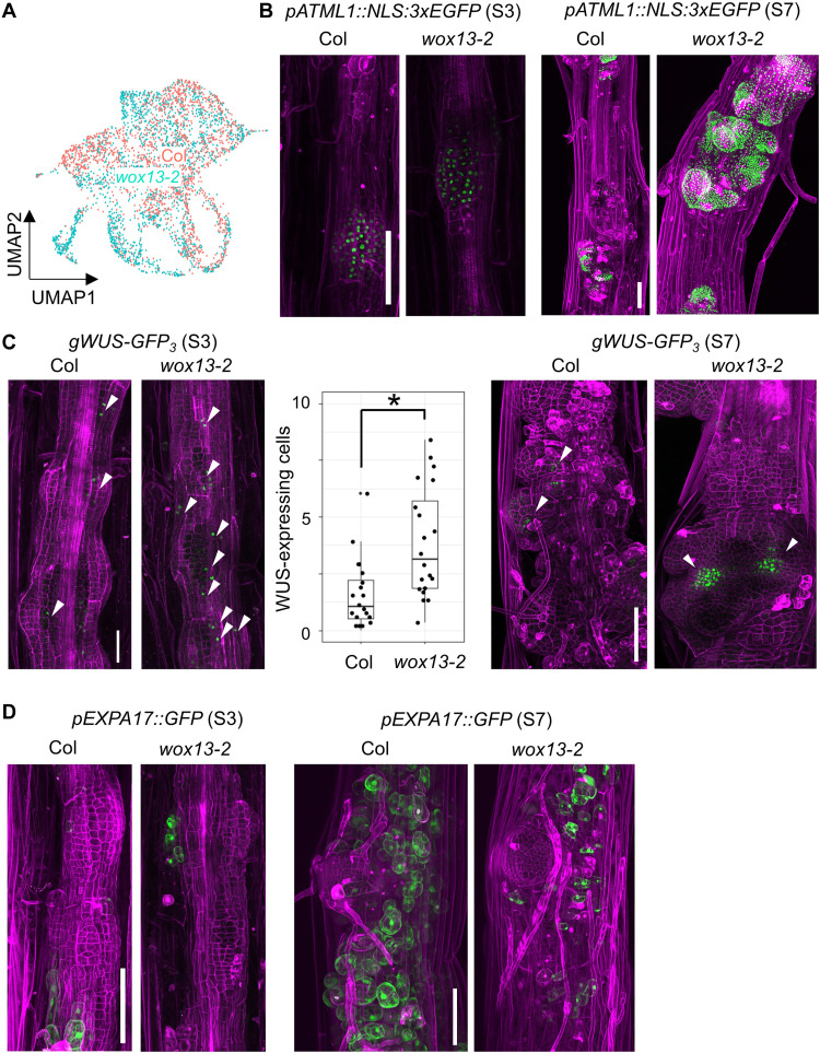
Plants can regenerate their bodies via de novo establishment of shoot apical meristems (SAMs) from pluripotent callus. Only a small fraction of callus cells is eventually specified into SAMs but the molecular mechanisms underlying fate specification remain obscure. The expression of WUSCHEL (WUS) is an early hallmark of SAM fate acquisition. Here, we show that a WUS paralog, WUSCHEL-RELATED HOMEOBOX 13 (WOX13), negatively regulates SAM formation from callus in Arabidopsis thaliana. WOX13 promotes non-meristematic cell fate via transcriptional repression of WUS and other SAM regulators and activation of cell wall modifiers. Our Quartz-Seq2-based single cell transcriptome revealed that WOX13 plays key roles in determining cellular identity of callus cell population. We propose that reciprocal inhibition between WUS and WOX13 mediates critical cell fate determination in pluripotent cell population, which has a major impact on regeneration efficiency.


Figures

Fig. 1.
Fig. 1.. WOX13 suppresses shoot regeneration from pluripotent callus.
(A) Schematic diagram of tissue culture procedure. Hypocotyl explants from etiolated seedlings were incubated on CIM for 4 days and then transferred to SIM for further incubation. (B and C) Shoot regeneration phenotype of wox13-2 mutant and the complementing line (gWOX13-GFP in wox13-2). Representative images of explants (B) and quantitative data of shoot number per explant on SIM at day 11 (C). Alphabetical letters indicate statistical significance determined by analysis of variance (ANOVA) and Tukey’s multiple comparisons test (n ≥ 40, P < 0.05). (D) Time-series examination on shoot regeneration phenotype (n ≥ 2, 20 explants for each replicate). Shoot regeneration rate shows the frequency of explants that bear at least one shoot. (E) Representative images of explants on SIM at day 14. The inset shows the magnified image of the explant of Col and gWOX13-GFP, where highly expanded globular cells are present. (F) Spatiotemporal expression of gWOX13-GFP during tissue culture incubation. WOX13 expression is visualized by GFP (green) and cellular outlines are visualized by propidium iodide (PI) staining (magenta). Scale bars, 1 mm (B) and (E) and 0.1 mm (F).
Fig. 2.
Fig. 2.. Shoot meristem regulators and cell wall modifiers are misregulated in the wox13 mutant.
(A) Heatmap graphs showing the expression of genes involved in shoot meristem formation. Genes that display significantly different expression in at least one time point are highlighted by asterisks (edgeR, P < 0.05). (B) Representative examples of DEGs in (A) retrieved from RNA-seq dataset. Asterisks show significant difference at respective time points (edgeR, P < 0.05). The expression is shown in Transcripts Per Kilobase Million (TPM). (C) Heatmap graphs showing the expression of genes listed in the GO category “cell wall modification.” (D) Representative examples in (C) retrieved from RNA-seq dataset. Asterisks show significant difference at respective time points (edgeR, P < 0.05). Time course expression data were obtained on day 0 (0), CIM at day 4 (C4), SIM at day 4 (S4), and SIM at day 7 (S7).
Fig. 3.
Fig. 3.. WOX13 suppresses the expression of shoot meristem regulators and induces cell wall modifiers.
(A and B) Shoot regeneration phenotype of gWOX13-GR with DEX (DEX+) or without DEX (DEX) application during tissue culture. Scale bar, 1 mm. Representative images of explants (A) and quantitative data of shoot number per explant on SIM at day 11 (B). (C and D) Reverse transcription quantitative polymerase chain reaction analysis of shoot meristem regulators (C) and cell wall modifiers (D) upon WOX13 induction by DEX application. gWOX13-GR hypocotyl explants were transferred to +DEX or −DEX SIM media on SIM at day 3 and analyzed at 6 or 24 hours after the transfer. Data are means ± SE (n = 4, biological replicates). Asterisks indicate significant differences based on t test (P < 0.05).
Fig. 4.
Fig. 4.. scRNA-seq revealed the composition of heterogeneous callus cell population.
(A) UMAP plot of the scRNA-seq dataset analyzing 3987 isolated protoplast cells from SIM at day 7. Clusters representing SAM, LP (leaf primordia), highly expanded globular cells, and proliferating cells in S phase and G2-M phase are annotated. (B) Dot blot visualization of genes listed as markers of clusters. (C) Reporter GFP expression of each gene in callus undergoing meristem initiation on SIM at day 7. Arrowheads indicate promeristems or SAMs. Red circles highlight specific cluster(s) for gene expression. Scale bar, 0.1 mm. (D) A schematic illustration of the spatial expression pattern of marker genes.
Fig. 5.
Fig. 5.. Differential callus cell composition between WT and the wox13 mutant.
(A) UMAP plot of the scRNA-seq dataset describing the differential cellular composition of callus cells depending on genotypes. (B to D) Reporter GFP expression of ATML1 (B), WUS (C), and EXPA17 (D) on SIM at day 3 (S3) or SIM at day 7 (S7). Arrowheads indicate GFP-positive cells. Scale bars, 0.1 mm.
Fig. 6.
Fig. 6.. Spatial separation of WOX13 and WUS expression domains.
(A and B) Live imaging of gWOX13-mRUBY (magenta) and gWUS-GFP3 (green). Representative cases from two types of relative expression pattern are shown. Consecutive observation was performed daily from SIM at day 6 (S6) to SIM at day 8 (S8) or SIM at day 9 (S9). (C and D) Snapshot image of gWOX13-mRUBY (magenta) and gWUS-GFP3 (green) in promeristems (C) and SAM (D). Asterisks show leaf primordia. Scale bars, 50 μm. (E) A schematic illustration of the spatial expression pattern of WOX13 and WUS.
Fig. 7.
Fig. 7.. Mutually repressive WOX13 and WUS demarcate callus cell identity.
(A and B) Reporter luciferase assay to analyze WUS promoter activity upon WOX13 overexpression (A) and WOX13 promoter activity upon WUS overexpression (B). Data are means ± SE [n = 9 in (A) and n = 3 in (B), biological replicates]. (C) ChIP-seq data showing WUS-mCherry binding to WOX13 locus. The data are retrieved from a previous report (33). (D) Genetic interaction between wox13 and wus. Representative image of the explant on SIM at day 21 are shown. Scale bar, 1 mm. (E) A schematic diagram describing WOX13 function in the control of callus cell fate and its relationships with WUS. The top panel summarizes the regulatory scheme, and the bottom panel describes the spatiotemporal expression pattern of WOX13 (magenta) and WUS (green).

References

    1. K. D. Birnbaum, A. S. Alvarado, Slicing across kingdoms: Regeneration in plants and animals. Cell 132, 697–710 (2008). - PMC - PubMed
    1. M. Ikeuchi, D. S. Favero, Y. Sakamoto, A. Iwase, D. Coleman, B. Rymen, K. Sugimoto, Molecular mechanisms of plant regeneration. Annu. Rev. Plant Biol. 70, 377–406 (2019). - PubMed
    1. D. Valvekens, M. Van Montagu, M. Van Lijsebettens, Agrobacterium tumefaciens-mediated transformation of Arabidopsis thaliana root explants by using kanamycin selection. Proc. Natl. Acad. Sci. U.S.A. 85, 5536–5540 (1988). - PMC - PubMed
    1. K. Sugimoto, Y. Jiao, E. M. Meyerowitz, Arabidopsis regeneration from multiple tissues occurs via a root development pathway. Dev. Cell 18, 463–471 (2010). - PubMed
    1. A. Kareem, K. Durgaprasad, S. Sugimoto, Y. Du, A. J. Pulianmackal, Z. B. Trivedi, P. V. Abhayadev, V. Pinon, E. M. Meyerowitz, B. Scheres, K. Prasad, PLETHORA genes control regeneration by a two-step mechanism. Curr. Biol. 25, 1017–1030 (2015). - PMC - PubMed

MeSH terms