. 2023 Jul 7;9(27):eadg4846.
doi: 10.1126/sciadv.adg4846. Epub 2023 Jul 7.

Essential autoproteolysis of bacterial anti-σ factor RsgI for transmembrane signal transduction

Affiliations

Essential autoproteolysis of bacterial anti-σ factor RsgI for transmembrane signal transduction

Chao Chen et al. Sci Adv. .

Abstract

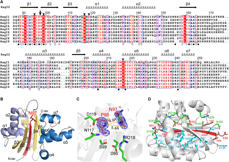
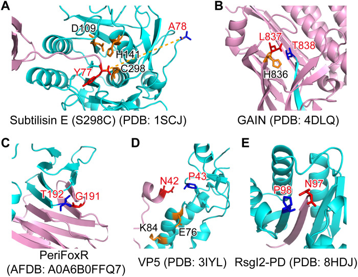
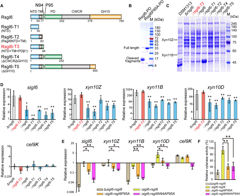
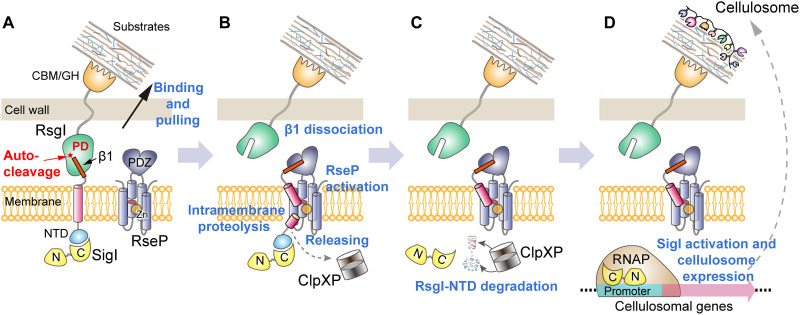
Autoproteolysis has been discovered to play key roles in various biological processes, but functional autoproteolysis has been rarely reported for transmembrane signaling in prokaryotes. In this study, an autoproteolytic effect was discovered in the conserved periplasmic domain of anti-σ factor RsgIs from Clostridium thermocellum, which was found to transmit extracellular polysaccharide-sensing signals into cells for regulation of the cellulosome system, a polysaccharide-degrading multienzyme complex. Crystal and NMR structures of periplasmic domains from three RsgIs demonstrated that they are different from all known proteins that undergo autoproteolysis. The RsgI-based autocleavage site was located at a conserved Asn-Pro motif between the β1 and β2 strands in the periplasmic domain. This cleavage was demonstrated to be essential for subsequent regulated intramembrane proteolysis to activate the cognate SigI, in a manner similar to that of autoproteolysis-dependent activation of eukaryotic adhesion G protein-coupled receptors. These results indicate the presence of a unique prevalent type of autoproteolytic phenomenon in bacteria for signal transduction.


Figures

Fig. 1.
Fig. 1.. Identification of the autocleavage phenomenon and the cleavage site of C. thermocellum RsgI2.
(A) Schematic representation of the composition and the location of the autocleavage site of RsgI2. RsgI2 contains an NTD, a transmembrane helix (TM), a PD, a probable cell wall–crossing region (CWCR) and a C-terminal substrate-binding CBM3b domain (CBM). The amino acid sequence around the autocleavage site is shown, and the first five amino acid residues identified by N-terminal protein sequencing are colored in red. (B) Western-blot analysis of RsgI2 expressed in C. thermocellum. The mutant ΔrsgI2 (control), the ΔrsgI2 bearing the plasmids to express RsgI2 with either an N- or C-terminal HA-tag (RsgI2-NHA and RsgI2-CHA), and the P98A mutant RsgI2P98A-NHA were cultured on the different designated carbon sources, and the resultant labeled RsgI2 components were detected by an anti–HA-tag antibody. (C) SDS–polyacrylamide gel electrophoresis (SDS-PAGE) analysis of the purified SMT3-RsgI2-PD protein and its N97A and P98A mutants. The results of N-terminal protein sequencing for the two bands of SMT3-RsgI2-PD are denoted parenthetically on the left. (D) RsgI2-PD fused with an N-terminal glutathione S-transferase (GST) and a C-terminal FLAG tag (RsgI2-PD) and its P98A mutant (RsgI2P98A-PD) were expressed using the PURE cell-free transcription/translation system and analyzed by Western blotting using an anti-FLAG antibody.
Fig. 2.
Fig. 2.. Structure of RsgI2-PD.
(A) Sequence alignments of the RsgI-PDs from C. thermocellum. The autocleavage site and the surrounding conserved residues are indicated by an arrow and filled triangles, respectively, at the top. The residues involved in the polar and hydrophobic interactions between the disconnected β1 and the other parts of the protein are indicated by filled and open circles, respectively, at the bottom. (B) Ribbon representation of the crystal structure of RsgI2-PD. The disconnected β1 caused by the autocleavage is colored in red, and residues Asn97 and Pro98 are shown as sticks. (C) The conserved residues around the Asn97 and Pro98. The 2mFo-DFc densities for Asn97 and Pro98 are contoured in blue at 1.0σ. The mFo-DFc maps are shown in green and red for positive and negative densities, respectively, at 3.0σ. (D) The interactions of β1 with other parts of the protein. The residues of α helix subdomains involved in the polar and hydrophobic interactions are shown as green and cyan sticks, respectively.
Fig. 3.
Fig. 3.. The structure of RsgI-PD is distinct from other known autocleaved proteins.
The N- and C-terminal parts are colored pink and cyan, respectively. The P − 1 and P + 1 residues of the cleavage site are shown in red and blue sticks, respectively, and other residues probably involved in the catalysis are shown in orange sticks. (A) The structure of the propeptide-subtilisin E (S298C mutant) complex from B. subtilis. (B) The crystal structure of the GAIN domain of the GPCR CL1. (C) The AlphaFold2 structural model of the PD of FoxR (PeriFoxR) from P. aeruginosa. (D) The structure of the VP5 protein of Aquareovirus. (E) The structure of RsgI2-PD from C. thermocellum.
Fig. 4.
Fig. 4.. Analysis of SigI6 and xylanase expression in mutant strains to demonstrate the role of autoproteolysis in signaling.
(A) Schematic representation of the composition of RsgI6 and its truncation mutants. RsgI6 contains an intracellular NTD, a TM, a PD, a proposed CWCR, and a C-terminal substrate-binding GH10 domain (GH10). (B) SDS-PAGE analysis of the purified SMT3-RsgI6-PD protein and its P95A mutant. (C) SDS-PAGE analysis of the extracellular proteins of wild type (DSM1313), ΔrsgI6, and its complemented strains with various RsgI6 truncations of C. thermocellum DSM1313, cultured in media with cellobiose as the carbon source. (D) qRT-PCR analysis of the genes of SigI6 and cellulosomal xylanases in ΔrsgI6 and its complemented strains grown in the cellobiose medium. The relative expression represents the ratio of transcribed mRNA levels in the mutant strains compared to that of DSM1313. The P values were calculated on the basis of three replicates using Student’s t test with ΔrsgI6::rsgI6-T3 as the reference. (E) qRT-PCR analysis of ΔsigI6-rsgI6 and its complemented strains grown in medium with alkali-pretreated wheat straw as carbon source. The relative expression represents the ratio of transcribed mRNA levels in the mutant strains compared to that of DSM1313. (F) Xylanase activity of the culture supernatant. The P values in (D) and (E) were calculated on the basis of three replicates using Student’s t test. *P < 0.05 and **P < 0.01.
Fig. 5.
Fig. 5.. The role of RseP and ClpXP in the signal transduction of RsgI6.
(A) qRT-PCR analysis of SigI6 and xylanase expression in ΔrsgI6, ΔrsgI6ΔrseP, and their rsgI6-T3-complemented strains. The relative expression represents the ratio of the mRNA levels in the mutant strains compared to that of the wild-type DSM1313 strain. The P values were calculated on the basis of three replicates using Student’s t test between ΔrsgI6ΔrseP and ΔrsgI6ΔrseP::rsgI6-T3. *P < 0.05 and **P < 0.01. (B) SDS-PAGE analysis of the extracellular proteins of ΔrsgI6, ΔrsgI6ΔrseP, and their rsgI6-T3–complemented strains cultured with cellobiose as the carbon source. (C) Sequence alignment of the transmembrane helix regions of the RsgIs in C. thermocellum. The transmembrane helices predicted by TMHMM (65) are indicated by blue rectangles. A consensus WebLogo (66) is shown at the bottom. The red vertical line indicates the Asn-Pro cleavage site. The potential ClpP recognition motifs are colored in red. (D) Protease assay using RsgI6NTD-VAA1 as the substrate. The positions of the proteases and substrates in the control (the mixture before the reaction) are indicated by arrows. The degradation of substrate bands is indicated by a red dashed rectangle.
Fig. 6.
Fig. 6.. Proposed model of transmembrane signal transduction of RsgI in C. thermocellum.
(A) Autocleavage of the conserved Asn-Pro motif and binding of the extracellular RsgI-based CBM to substrate. (B) Recognition and cleavage of the β1 fragment and the transmembrane helix by the RseP protease; release of the NTD. (C) Degradation of the NTD by the cytosolic protease ClpXP. (D) Release of SigI and transcription of cellulosomal genes.

Similar articles

Cited by

References

    1. S. C. Jain, U. Shinde, Y. Li, M. Inouye, H. M. Berman, The crystal structure of an autoprocessed Ser221Cys-subtilisin E-propeptide complex at 2.0 Å resolution. J. Mol. Biol. 284, 137–144 (1998). - PubMed
    1. H. Ikemura, M. Inouye, In vitro processing of pro-subtilisin produced in Escherichia coli. J. Biol. Chem. 263, 12959–12963 (1988). - PubMed
    1. A. Vizurraga, R. Adhikari, J. Yeung, M. Yu, G. G. Tall, Mechanisms of adhesion G protein-coupled receptor activation. J. Biol. Chem. 295, 14065–14083 (2020). - PMC - PubMed
    1. K. C. Bastiaansen, J. R. Otero-Asman, J. Luirink, W. Bitter, M. A. Llamas, Processing of cell-surface signalling anti-sigma factors prior to signal recognition is a conserved autoproteolytic mechanism that produces two functional domains. Environ. Microbiol. 17, 3263–3277 (2015). - PubMed
    1. K. C. Bastiaansen, P. van Ulsen, M. Wijtmans, W. Bitter, M. A. Llamas, Self-cleavage of the Pseudomonas aeruginosa cell-surface signaling anti-sigma factor FoxR occurs through an N-O acyl rearrangement. J. Biol. Chem. 290, 12237–12246 (2015). - PMC - PubMed

MeSH terms