Review
. 2023 Jul 14;8(85):eabq3015.
doi: 10.1126/sciimmunol.abq3015. Epub 2023 Jul 7.

The interplay between the DNA damage response and ectonucleotidases modulates tumor response to therapy

Affiliations
Review

The interplay between the DNA damage response and ectonucleotidases modulates tumor response to therapy

John Stagg et al. Sci Immunol. .

Abstract

The extracellular nucleoside adenosine reduces tissue inflammation and is generated by irreversible dephosphorylation of adenosine monophosphate (AMP) mediated by the ectonucleotidase CD73. The pro-inflammatory nucleotides adenosine triphosphate, nicotinamide adenine dinucleotide, and cyclic guanosine -monophosphate-AMP (cGAMP), which are produced in the tumor microenvironment (TME) during therapy-induced immunogenic cell death and activation of innate immune signaling, can be converted into AMP by ectonucleotidases CD39, CD38, and CD203a/ENPP1. Thus, ectonucleotidases shape the TME by converting immune-activating signals into an immunosuppressive one. Ectonucleotidases also hinder the ability of therapies including radiation therapy, which enhance the release of pro-inflammatory nucleotides in the extracellular milieu, to induce immune-mediated tumor rejection. Here, we review the immunosuppressive effects of adenosine and the role of different ectonucleotidases in modulating antitumor immune responses. We discuss emerging opportunities to target adenosine generation and/or its ability to signal via adenosine receptors expressed by immune and cancer cells in the context of combination immunotherapy and radiotherapy.


Conflict of interest statement

Author contributions

All authors wrote the manuscript.

Competing interests

SD has received compensation for consultant/advisory services from Lytix Biopharma, Johnson & Johnson Enterprise Innovation Inc., EMD Serono, Ono Pharmaceutical, and Genentech, and research support from Lytix Biopharma and Boehringer-Ingelheim for unrelated projects.

JS owns stock of Surface Oncology and has received compensation for consultant/advisory services from Surface Oncology and Domain Therapeutics, and research support from Surface Oncology and Domain Therapeutics for unrelated projects. EG receives research support from Arcus Biosciences, Inc. for the PANTHER trial.

The authors declare that they have no competing interests related to this work.

Figures

Figure 1.
Figure 1.. The immunogenicity of radiotherapy is regulated by ectonucleotidases.
1. Radiotherapy -induced cytosolic DNA leads to activation of cGAS that generates cGAMP, part of which can be exported into the extracellular compartment where it activates STING in immune cells. Downstream IFN-I promotes cross-presenting dendritic cell (DC) activation and priming of anti-tumour T cells. 2. ENPP1 converts cGAMP into AMP, generating a substrate for adenosine generation by CD73. 3. ATP released by irradiated cancer cells binds to P2X7R on DCs, promoting production of pro-inflammatory cytokines. 4. CD39 converts ATP into ADP and AMP which is then converted into adenosine by CD73. Adenosine exerts immune suppression by binding to adenosine receptors A2AR and A2BR on immune cells. 5. Extracellular NAD+ also contributes to adenosine generation by sequential conversion into ADPR mediated by CD38, into AMP mediated by ENPP1, and into adenosine via CD73. In addition, NAD+ serves as a substrate for ADP-ribosyltransferases (ARTs) including ART1 which mono-ADP-ribosylate (MARylate) the P2X7R on T cells to induce NAD-induced cell death (NICD). Importantly CD38 and CD39 expressed by immune cells can offset NICD and ATP-induced cell death by catabolizing NAD+ and ATP respectively, thus providing cytoprotective roles.
Figure 2.
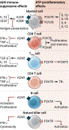
Figure 2.. Immune regulation through extracellular adenosine (ADO) and adenosine triphosphate (ATP).
In the tumor microenvironment, ADO activates A2AR and A2BR to promote the production of IL-10 and VEGF by myeloid cells, thereby suppressing antigen presentation. In contrast, ATP activates P2X7R and P2Y2R to enhance production of proinflammatory IL-1β, IL-18, IL-6 and IL-12. In CD8+ effector T cells (CD8), A2AR inhibits IFN-γ, TNF-α and proliferation, while P2X7R and P2X4R promote T cell proliferation and immune memory. A2AR further enhances the suppressive function of T regulatory cells (Treg), while P2X7R inhibits FOXP3 expression. In CD4+ T effector cells (CD4), A2AR inhibits IFN-γ, TNF-α and proliferation, while activation of P2X7R promotes T follicular helper (Tfh) function. ADO inhibits B cells via A2AR, while P2X7R promotes B cell activation. Finally, activation of A2AR and/or P2X7R in natural killer (NK) cells inhibit their function.
Figure 3.
Figure 3.. Crosstalk between DNA damage and adenosine signaling.
DNA damage induces upregulation of CD73 and CD39 ectonucleotidases via CREB and HIF-1. Adenosine produced by CD39 and CD73 signals through A2BR leading to the activation of PKA, PKC and ERK pathways, which have been associated with increased DNA damage responses (DDR). DNA damage also promotes TGF-β signaling, which increases ectonucleotidase expression. EMT enhances DDR and also upregulate CD73, which in turn supports EMT via A2BR signaling.
Figure 4.
Figure 4.. Ectonucleotidases inhibit the cGAS-STING pathway.
Upon DNA damage, the cytosolic sensor cGAS produces cyclic GMP-AMP (cGAMP), thereby activating STING and the transcription of type I IFNs and pro-inflammatory cytokines via IRF3 and NF-kB. Cytosolic cGAMP is also released via transporters and gap junctions to activate STING in neighbouring cells. Paracrine activation of STING, notably in antigen presenting cells (APC) and cancer cells, is suppressed by ENPP1 that hydrolyses cGAMP into AMP and GMP. ENPP1 and CD39 further hydrolyses extracellular ATP into AMP. Extracellular AMP is also produced from the conversion of NAD+ to ADPR by CD38, followed by ADPR hydrolysis by ENPP1. Extracellular AMP is hydrolyzed by CD73 into adenosine (ADO), activating cAMP-elevating A2AR and A2BR, which suppresses inflammatory responses, including by blocking NF-kB activity.

Similar articles

Cited by

References

    1. Sharma P, Allison JP, The future of immune checkpoint therapy. Science 348, 56–61 (2015). - PubMed
    1. Sharma P, Hu-Lieskovan S, Wargo JA, Ribas A, Primary, Adaptive, and Acquired Resistance to Cancer Immunotherapy. Cell 168, 707–723 (2017). - PMC - PubMed
    1. Charpentier M, Spada S, Van Nest SJ, Demaria S, Radiation therapy-induced remodeling of the tumor immune microenvironment. Semin Cancer Biol, (2022). - PubMed
    1. Ghiringhelli F, Apetoh L, Tesniere A, Aymeric L, Ma Y, Ortiz C, Vermaelen K, Panaretakis T, Mignot G, Ullrich E, Perfettini JL, Schlemmer F, Tasdemir E, Uhl M, Genin P, Civas A, Ryffel B, Kanellopoulos J, Tschopp J, Andre F, Lidereau R, McLaughlin NM, Haynes NM, Smyth MJ, Kroemer G, Zitvogel L, Activation of the NLRP3 inflammasome in dendritic cells induces IL-1beta-dependent adaptive immunity against tumors. Nat Med 15, 1170–1178 (2009). - PubMed
    1. Golden EB, Frances D, Pellicciotta I, Demaria S, Helen Barcellos-Hoff M, Formenti SC, Radiation fosters dose-dependent and chemotherapy-induced immunogenic cell death. Oncoimmunology 3, e28518 (2014). - PMC - PubMed