. 2023 Jul 26;145(29):15796-15808.
doi: 10.1021/jacs.3c02313. Epub 2023 Jul 7.

Serial Femtosecond Crystallography Reveals that Photoactivation in a Fluorescent Protein Proceeds via the Hula Twist Mechanism

Affiliations

Serial Femtosecond Crystallography Reveals that Photoactivation in a Fluorescent Protein Proceeds via the Hula Twist Mechanism

Alisia Fadini et al. J Am Chem Soc. .

Abstract

Chromophore cis/trans photoisomerization is a fundamental process in chemistry and in the activation of many photosensitive proteins. A major task is understanding the effect of the protein environment on the efficiency and direction of this reaction compared to what is observed in the gas and solution phases. In this study, we set out to visualize the hula twist (HT) mechanism in a fluorescent protein, which is hypothesized to be the preferred mechanism in a spatially constrained binding pocket. We use a chlorine substituent to break the twofold symmetry of the embedded phenolic group of the chromophore and unambiguously identify the HT primary photoproduct. Through serial femtosecond crystallography, we then track the photoreaction from femtoseconds to the microsecond regime. We observe signals for the photoisomerization of the chromophore as early as 300 fs, obtaining the first experimental structural evidence of the HT mechanism in a protein on its femtosecond-to-picosecond timescale. We are then able to follow how chromophore isomerization and twisting lead to secondary structure rearrangements of the protein β-barrel across the time window of our measurements.


Conflict of interest statement

The authors declare no competing financial interest.

Figures

Figure 1
Figure 1
Schematic of potential chromophore photoisomerization pathways. Two possible pathways for the cis/trans photoisomerization of a chromophore are the OBF pathway or the HT pathway, both shown here schematically. The OBF mechanism involves the rotation of only the isomerizing bond, τ, and is expected to sweep a large volume, as half of the molecule is flipped in the process. In the HT pathway, on the other hand, both τ and its neighboring bond ϕ rotate simultaneously. The products formed by these two pathways are indistinguishable if the part of the molecule that flips is symmetric.
Figure 2
Figure 2
Cl-rsEGFP2 photoswitching. (a) Reversibly photoswitchable protein rsEGFP2 can be converted between a dark OFF state to a fluorescent ON state by specific frequencies of light. These changes are caused by a trans-to-cis isomerization and subsequent deprotonation of its embedded chromophore. (b) Addition of a chlorine substituent to the phenolate ring of the chromophore can break its C2 point group symmetry and allow us to distinguish between the HT and OBF mechanisms of chromophore photoisomerization in the first photoproduct of the OFF-to-ON reaction. The normalized absorption spectrum and structure of Cl-rsEGFP2 (c) possess very similar properties as the non-chlorinated protein: the OFF state absorbing predominantly at 400 nm and the ON state at around 480 nm, while the protein tertiary structure exhibits the β-barrel fold typical of GFP-like constructs. A 2 nm shift can be observed in the absorption profile between chlorinated and non-chlorinated constructs caused by the electron-withdrawing nature of chlorine.
Figure 3
Figure 3
OFF-state chromophore configurations in Cl-rsEGFP2. (a) Refined Cl-rsEGFP2 dark OFF-state structure obtained from room-temperature serial femtosecond crystallography (PDB 8A6G). The predominant chromophore (OHD) conformation is the “planar” trans anti (trans-PL). Minor populations of a “twisted” trans syn (trans-TW) and of cis anti configurations are also modeled (refined occupancies of 12 and 14%, respectively). The three chromophore configurations are accompanied by three alternate His149 conformations. Two Val151 conformations are also resolved and matched to the trans-PL and trans-TW species. (b) OFF-state equilibrium trans structures of Cl-rsEGFP2 and (c) rsEGFP2. The major configuration found in the OFF-state chlorinated structure is trans-PL, while it is trans-TW when the chlorine substituent is not present. We presume that the heavy atom substitution results in a higher energy for the trans-TW state through increased steric hindrance. See also Figures S4–S6, Table S3, and Note S1.
Figure 4
Figure 4
Time-resolved DED maps of Cl-rsEGFP2. Q-weighted DED maps for the collected pump–probe delays. Positive (blue) and negative (red) DED is shown at ±3.5 σ. The refined trans anti and cis anti species are shown in light and dark gray, respectively. In the 600 fs, 900 fs, and 5 ps maps, the feature “Peak 1″ is highlighted: this is the main indicator of the presence of a femtosecond intermediate, which we have called trans-FS and discussed with further details in the main text. PDBs (in order) 8A6N, 8A6O, 8A6P, 8A6Q, 8A6R, and 8A6S. See also Figures S7–S13.
Figure 5
Figure 5
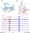
Light-induced changes across the entire Cl-rsEGFP2 protein structure. (a) The background-subtracted map (WΔFmax) for the 100 ps dataset clearly outlines the presence of the cis anti photoproduct. (b,c) The strongest signals in the Q-weighted DED maps for the collected time points are concentrated on the chromophore (OHD). However, by moving a spherical volume through all the atoms in the protein and integrating the negative and positive electron density within it, two other regions of variation stand out: the central α-helix (residues 58–67, orange) and the β-strand 7 (residues 146–152, blue). Signals in the α-helix are strongest in the early data points (c) and suggest a downward shift of the helix [(b) and Figure S15]. Negative density signals on β-strand 7 (Figure S16) are strongest on the late picosecond–microsecond timescale and suggest an increase in conformational flexibility for this secondary structure element. See also Figures S9–16.
Figure 6
Figure 6
Femtosecond Cl-rsEGFP2 TA data. (a) Transient difference absorption spectra recorded at different pump–probe time delays after a femtosecond laser excitation (400 nm) starting from the Cl-rsEGFP2 OFF state. (b) Components fitted through global analysis of the data shown in (a). (c) Raw concentration profiles for the four components globally fitted using a sequential model. See also Figures S17 and S18.
Figure 7
Figure 7
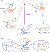
Comparison between experimental and calculated chromophore structures. (a) Molecular structures obtained through QM-MM modeling before and after CI crossing are shown with their energy arrangement relative to the Franck–Condon point and their respective torsion angles. Relative energy values computed at a lower (SA2-CASSCF(12,11)/3-21G//Amber03) and higher (XMCQDPT2/SA6-CASSCF(12,11)/cc-pVDZ//Amber03—in parenthesis) level of theory are stated (see also Section 4 in the Supporting Information Procedures). The photoexcited trans-PL species isomerizes in the protein binding pocket to the planar cis anti via HT, passing through a highly twisted conformation near the CI (trans-TWCI). An excited-state twisted minimum structure (trans-TWMIN) and a fluorescent trans minimum (trans-FL) are also found. (b) The calculated conformations are compared to the planar trans anti and cis anti species, as well as to the twisted trans-FS intermediate, refined from TR-SFX data. See also Figure S19.

References

    1. Gozem S.; Luk H. L.; Schapiro I.; Olivucci M. Theory and Simulation of the Ultrafast Double-Bond Isomerization of Biological Chromophores. Chem. Rev. 2017, 117, 13502–13565. 10.1021/acs.chemrev.7b00177. - DOI - PubMed
    1. Kandori H. Retinal Proteins: Photochemistry and Optogenetics. Bull. Chem. Soc. Jpn. 2020, 93, 76–85. 10.1246/bcsj.20190292. - DOI
    1. Zhou X. X.; Lin M. Z. Photoswitchable fluorescent proteins: ten years of colorful chemistry and exciting applications. Curr. Opin. Chem. Biol. 2013, 17, 682–690. 10.1016/j.cbpa.2013.05.031. - DOI - PMC - PubMed
    1. Cheng M.-C.; Kathare P. K.; Paik I.; Huq E. Phytochrome Signaling Networks. Annu. Rev. Plant Biol. 2021, 72, 217–244. 10.1146/annurev-arplant-080620-024221. - DOI - PMC - PubMed
    1. Mishra K.; Fuenzalida-Werner J. P.; Pennacchietti F.; Janowski R.; Chmyrov A.; Huang Y.; Zakian C.; Klemm U.; Testa I.; Niessing D.; et al. Genetically encoded photo-switchable molecular sensors for optoacoustic and super-resolution imaging. Nat. Biotechnol. 2022, 40, 598–605. 10.1038/s41587-021-01100-5. - DOI - PMC - PubMed

Publication types