. 2023 Sep:35:101734.
doi: 10.1016/j.tranon.2023.101734. Epub 2023 Jul 5.

NPRA promotes fatty acid metabolism and proliferation of gastric cancer cells by binding to PPARα

Affiliations

NPRA promotes fatty acid metabolism and proliferation of gastric cancer cells by binding to PPARα

Tingting Cao et al. Transl Oncol. 2023 Sep.

Abstract

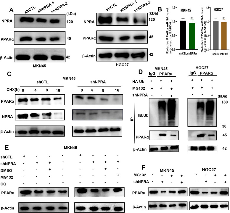
Among cancers, gastric cancer (GC) ranks third globally in morbidity and mortality, particularly in East Asia. Natriuretic peptide receptor A (NPRA), a receptor for guanylate cyclase, plays important roles in regulating water and sodium balance. Recent studies have suggested that NPRA is involved in tumorigenesis, but its role in GC development remains unclear. Herein, we showed that the expression level of NPRA was positively correlated with gastric tumor size and clinical stage. Patients with high NPRA expression had a lower five-year survival rate than those with low expression, and NPRA was identified as an independent predictor of GC prognosis. NPRA knockdown suppressed GC cell proliferation, migration and invasion. NPRA overexpression enhanced cell malignant behavior. Immunohistochemistry of collected tumor samples showed that tumors with high NPRA expression had higher peroxisome proliferator-activated receptor α (PPARα) levels. In vivo and in vitro studies showed that NPRA promotes fatty acid oxidation and tumor cell metastasis. Co-IP showed that NPRA binds to PPARα and prevents PPARα degradation. PPARα upregulation under NPRA protection activates arnitine palmitoyl transferase 1B (CPT1B) to promote fatty acid oxidation. In this study, new mechanisms by which NPRA promotes the development of GC and new regulatory mechanisms of PPARα were identified.

Keywords: CPT1B; EMT; FAO; NPRA; PPARα.


Conflict of interest statement

Declaration of Competing Interest The authors have no conflict of interest.

Figures

Fig 1
Fig. 1
The expression of NPRA was significantly increased in GC tissues compared to control tissues and positively correlated with the expression of PPARα. (A) IHC was used to detect the expression of NPRA and PPARα in GC tissues. (B) ROC time curves of NPRA. (C) Kaplan‒Meier overall survival (OS) curves were generated based on NPRA expression levels. (D) Organoids from two patients. (E) qRT‒PCR was used to analyze NPRA mRNA expression in GC tissues and corresponding adjacent noncancer tissues.
Fig 2
Fig. 2
Silencing NPRA can suppress the proliferation of GC cell lines and inhibit their migration and FAO.(A) MKN45 and HGC27 cell lines were transfected with shNPRA-1 and shNPRA-2 and a blank control vector (shCTL), respectively. the expression of NPRA was analyzed by qRT‒PCR. (B) Colony formation experiments showed that silencing NPRA significantly reduced the proliferation of GC cells. (C) The statistical results of B. (D) A CCK-8 assay was applied to detect the viability of GC cells. (E) EdU was used to detect cell proliferation. (F) The statistical results of E. (G) TCGA detaset analysis shows the positive correlations between NPRA and EMT markers. (H) The wound healing assay indicated that NPRA downregulation inhibited MKN45 cell migration. (I) The statistical results of H. (J) The statistical results of K. (K) Transwell migration assays showed that knockdown of NPRA reduced the cell migration ability; scale bar: 200 μm. (L) Silencing NPRA significantly increased the apoptosis rate of GC cells in the cytometry experiment. (M) Western blot analyses of EMT marker proteins in NPRA knockdown GC cells. (N) ATP content detection. (O) NPRA knockdown significantly suppressed FAO.
Fig 3
Fig. 3
Overexpression of NPRA enhances the proliferation, migration and FAO of GC cells. (A) Colony formation experiments showed that NPRA significantly enhanced the proliferation of GC cells. (B) CCK-8 was used to detect the viability of GC cells. (C) EdU was used to detect cell proliferation. (D) The wound healing assay indicated that NPRA upregulation enhanced MKN45 cell migration. (E) Transwell migration assays showed that increased NPRA expression enhanced GC cell migration ability. (F) Flow cytometry was used to detect cell apoptosis. (G) Western blotting was used to determine the levels of E-Cadherin, N-Cadherin, Snail and Vimentin. (H,I) NPRA overexpression significantly increased FAO and ATP values.
Fig 4
Fig. 4
PPARα is required for GC cell proliferation, migration and FAO. (A, B) Co-IP experiments in the GC cell lines MKN45 and HGC27showed that NPRA interacted with PPARα and vice versa. (C) CCK-8 assays showed that si-PPARα reduced the proliferation ability of GC cells. (D) EdU experiments showed that si-PPARα can reduce cell proliferation. (E) The statistical results of D. (F) Flow cytometry was used to detect cell apoptosis. (G) The statistical results of F. (H) Transwell assays showed that PPARα promoted the migration of GC cells. (I) The statistical results of H. (J, K) Wound healing assays showed that PPARα promoted the migration of GC cells. (L) The statistical results of J and K. (M, N) ATP and FAO levels in GC cells were significantly decreased by silencing PPARα.
Fig 5
Fig. 5
NPRA promotes proliferation, migration and FAO through PPARα. (A, B, C) EdU experiments, CCK-8 assays and colony formation assays were used to detect cell proliferation. (D) The statistical results of B. (E) The transfection of si-PPARα into NPRA-overexpressing cells significantly increased the apoptosis rate of GC cells according to the cytometry experiment. (F) The statistical results of E. (G) The application of siRNA to downregulate PPARα in NPRA-overexpressing cells significantly inhibited the invasion of GC cells compared with that of NPRA-overexpressing control siRNA cells. (H) The statistical results of G. (I) The application of siRNA to downregulate PPARα in NPRA-overexpressing cells significantly decreased wound closure rate of GC cells compared with that of NPRA-overexpressing control siRNA cells. (J) The statistical results of I. (K, L) The transfection of si-PPARα into NPRA-overexpressing cells reduced the ATP content and FAO of GC cells.
Fig 6
Fig. 6
NPRA regulates PPARα expression. (A) Western blot analysis showed that NPRA knockdown decreased PPARα expression. (B) The level of PPARα mRNA was not significantly altered in NPRA-silenced cells. (C) Western blot analysis of PPARα protein stability using CHX (an inhibitor of protein synthesis) when NPRA was knocked down. (D) The levels of ubiquitinated PPARα protein were increased in GC cells when NPRA was knocked in the presence of ubiquitin. (E) The proteasome inhibitor MG132 reversed the downregulation of PPARα protein induced by NPRA knockdown. (F) The reduction in PPARα protein by shNPRA was restored by MG132.
Fig 7
Fig. 7
NPRA promoted CPT1B expression and tumorigenesis in mice. (A) NPRA knockdown decreased PPARα and CPT1B expression. (B) PPARα knockdown decreased CPT1B expression. (C) By applying si-PPARα to NPRA-overexpressing cells, we found that PPARα and CPT1B expression obviously decreased. (D, E, F) NPRA knockdown MKN45 cells showed decreased tumor formation ability compared with control cells, as reflected by tumor size (D&E) and weight (F). (G) Schematic diagram of the role of NPRA in GC. In conclusion, NPRA activates CPT1B by binding PPARα, which promotes the interaction between NPRA and PPARα proteins in GC, stabilizes the PPARα protein, and promotes the downstream pathway and FAO.

References

    1. Sung H., et al. Global cancer statistics 2020: GLOBOCAN estimates of incidence and mortality worldwide for 36 cancers in 185 countries. CA Cancer J. Clin. 2021;71(3):209–249. - PubMed
    1. Allemani C., et al. Global surveillance of cancer survival 1995-2009: analysis of individual data for 25,676,887 patients from 279 population-based registries in 67 countries (CONCORD-2) Lancet. 2015;385(9972):977–1010. - PMC - PubMed
    1. Hanahan D., Weinberg R.A. Hallmarks of cancer: the next generation. Cell. 2011;144(5):646–674. - PubMed
    1. Li M., et al. Fatty acid oxidation: driver of lymph node metastasis. Cancer Cell Int. 2021;21(1):339. - PMC - PubMed
    1. Lee C.K., et al. Tumor metastasis to lymph nodes requires YAP-dependent metabolic adaptation. Science. 2019;363(6427):644–649. - PubMed