. 2023 Jul 6;30(7):950-961.e7.
doi: 10.1016/j.stem.2023.06.006.

Generating high-fidelity cochlear organoids from human pluripotent stem cells

Affiliations

Generating high-fidelity cochlear organoids from human pluripotent stem cells

Stephen T Moore et al. Cell Stem Cell. .

Abstract

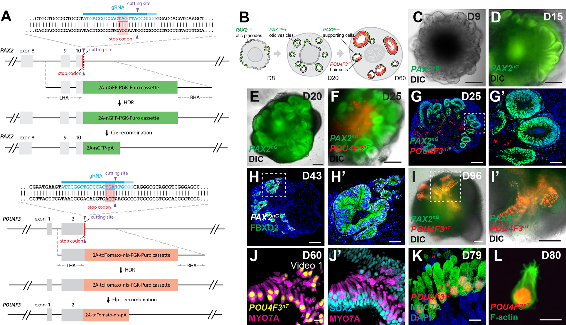
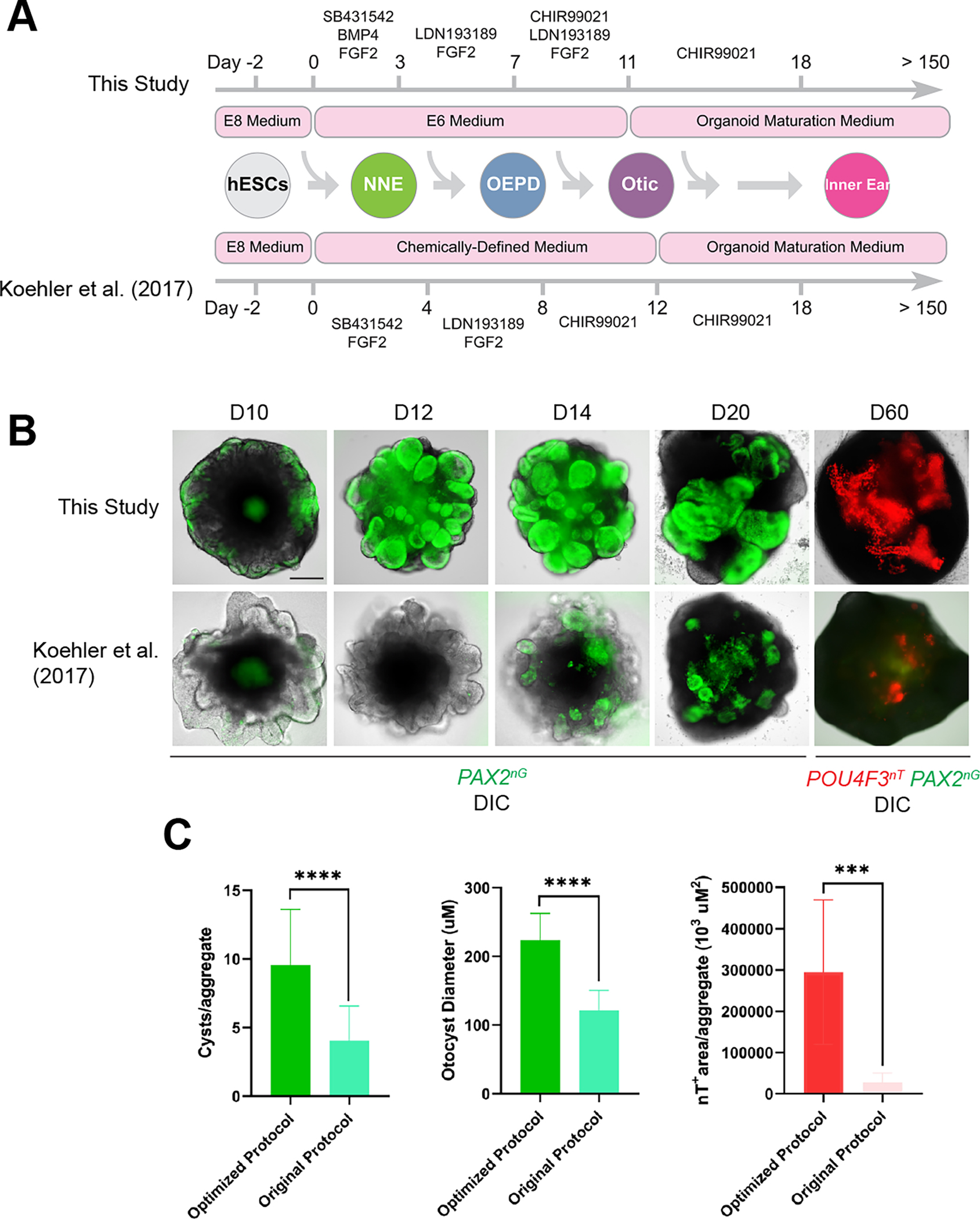
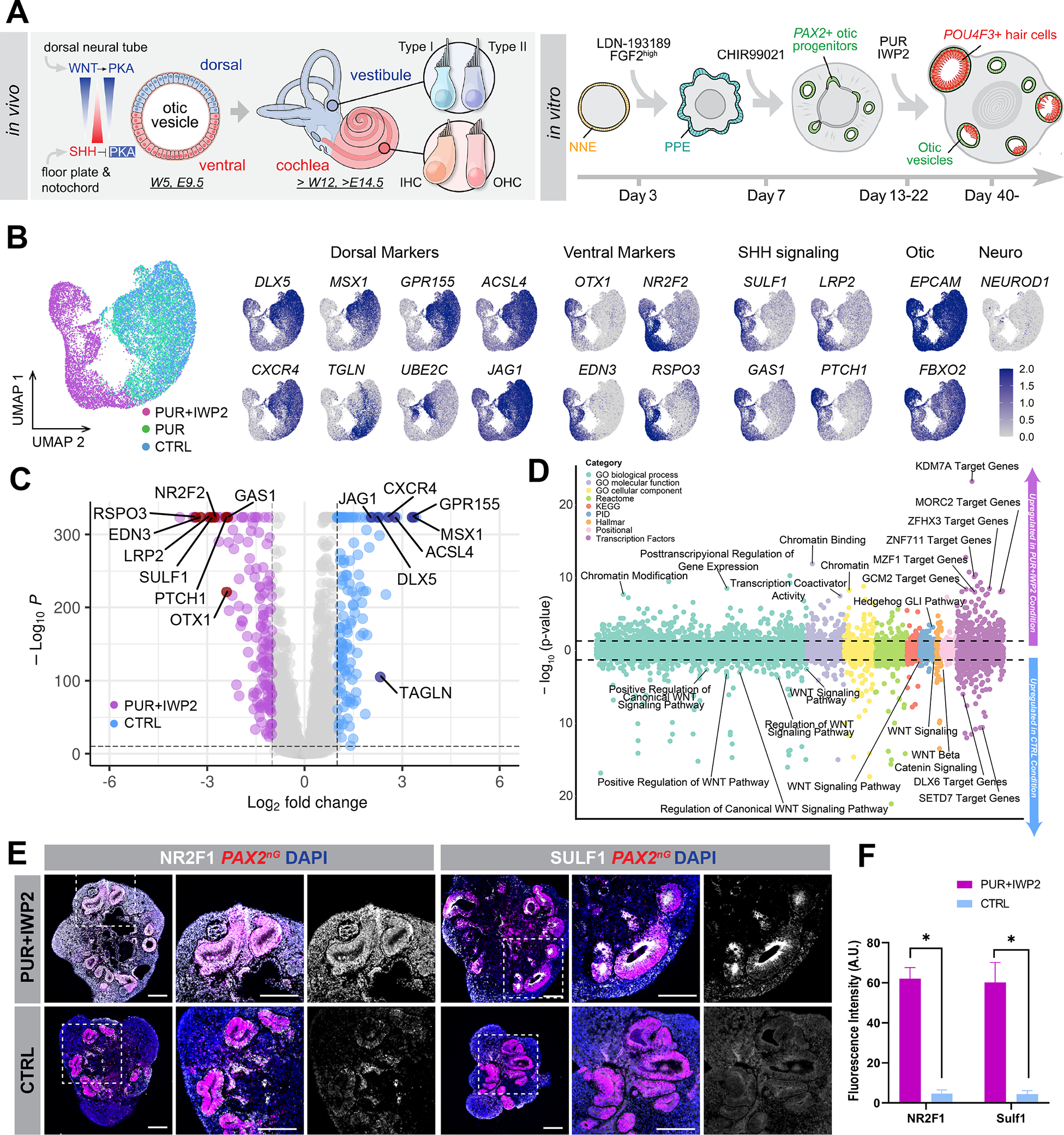
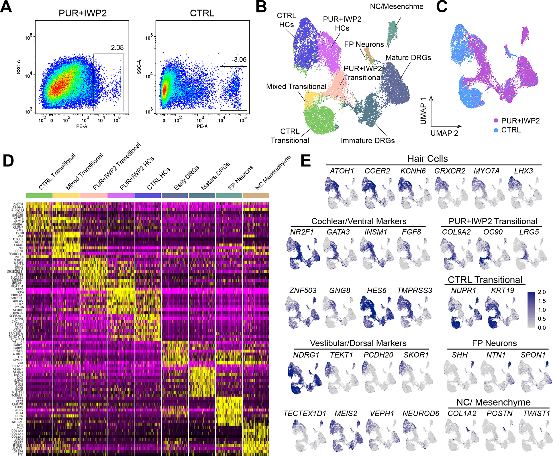
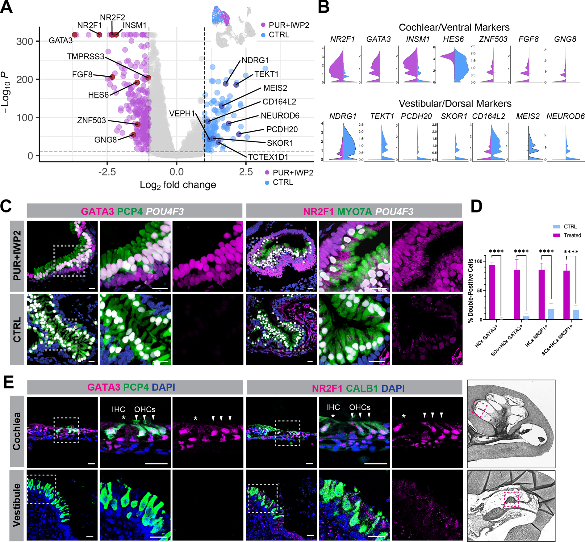
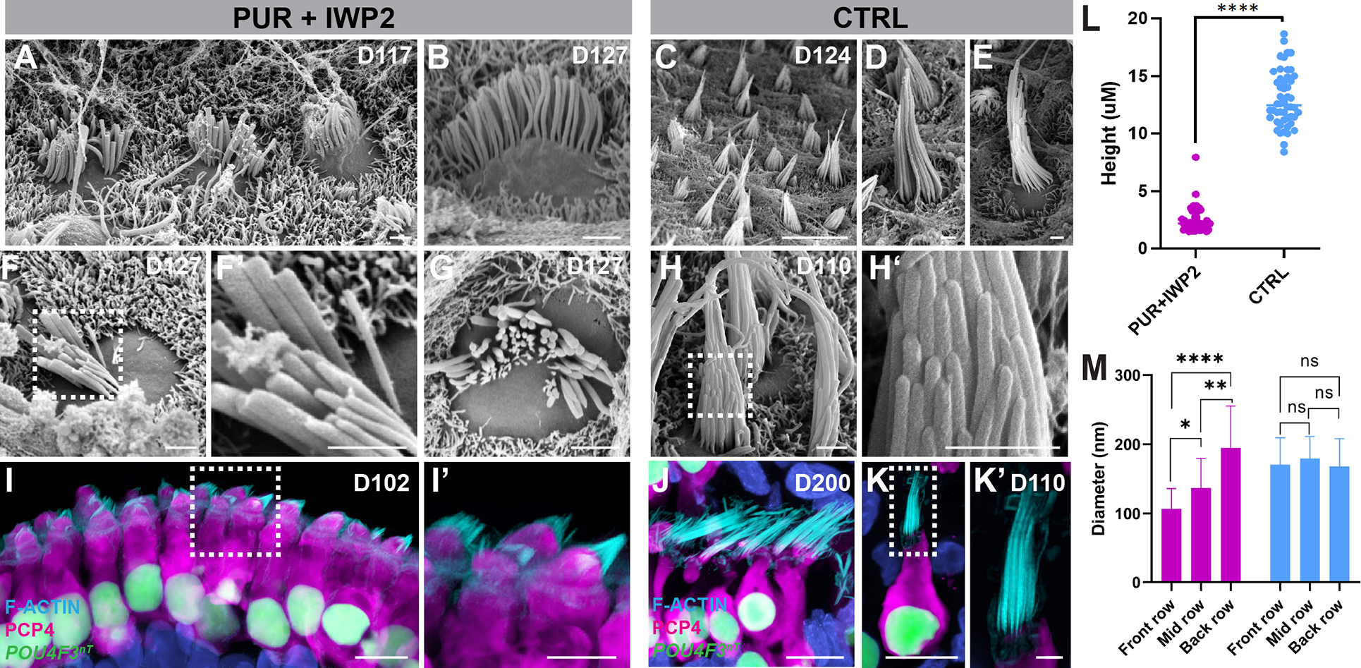
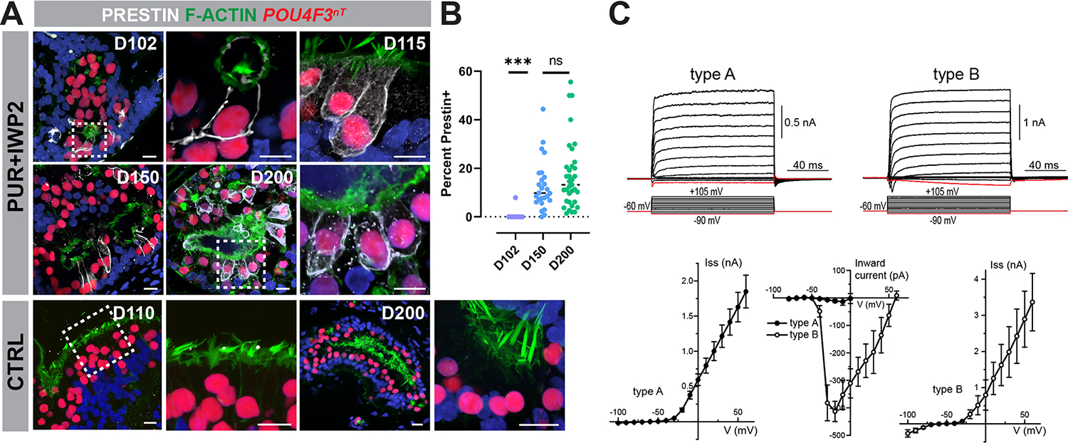
Mechanosensitive hair cells in the cochlea are responsible for hearing but are vulnerable to damage by genetic mutations and environmental insults. The paucity of human cochlear tissues makes it difficult to study cochlear hair cells. Organoids offer a compelling platform to study scarce tissues in vitro; however, derivation of cochlear cell types has proven non-trivial. Here, using 3D cultures of human pluripotent stem cells, we sought to replicate key differentiation cues of cochlear specification. We found that timed modulations of Sonic Hedgehog and WNT signaling promote ventral gene expression in otic progenitors. Ventralized otic progenitors subsequently give rise to elaborately patterned epithelia containing hair cells with morphology, marker expression, and functional properties consistent with both outer and inner hair cells in the cochlea. These results suggest that early morphogenic cues are sufficient to drive cochlear induction and establish an unprecedented system to model the human auditory organ.

Keywords: auditory; cochlea; hair cell; human; inner ear; organoid; pluripotent stem cell; scRNA-seq.


Conflict of interest statement

Declaration of interests S.T.M. and E.H. are the inventors on a PCT patent application titled “Methods of generating human cochlear hair cells.” The other authors declare no competing interests.

Figures

Figure 1.
Figure 1.. PAX2-2A-nGFP/POU4F3-2A-ntTomato (PAX2nG/POU4F3nT) multiplex reporter hESCs faithfully recapitulate otic progenitor and hair cell differentiations in inner ear organoids.
(A) Schematic illustrations of the PAX2-2A-nGFP and POU4F3-2A-ntdTomato CRISPR design. (B) Schematic of PAX2nG and POU4F3nT reporter expression during inner ear organoid development. (C-F) Live images of whole aggregates containing multiple developing inner ear organoids show the spatio-temporal progression of PAX2nG reporter expression and early morphogenesis of PAX2+ epithelium. (G-H) Representative images of hESC-derived aggregates showing PAX2nG+ epithelium organized into vesicles that co-express the otic-specific marker FBXO2, but devoid of POU4F3nT expression. (I-I’) Live images of late-stage (D96) aggregates showing intense POU4F3nT+ puncta localized to epithelial vesicles. (J-K) POU4F3nT+ cells in inner ear organoids also express the hair cell markers MYO7A and SOX2, and are located on the luminal surface of SOX2+ supporting epithelia. (L) Fixed cell suspension of dissociated POU4F3nT+ cells isolated from D80 inner ear organoids reveals tdTomato+ nuclei and F-actin+ membranes and stereocilia. Scale bars, 200 μm (C-G, H, I, I’), 50 μm (G’, H’), 10 μm (J-L).
Figure 2.
Figure 2.. Optimization of inner ear organoid derivation protocol.
(A) Schematic comparison of our original vs. optimized protocol. (B) Live images of whole cell aggregates containing inner ear organoids derived from our PAX2nG/POU4F3nT multiplex reporter hESC line cultured under the original vs. optimized protocol. (C) Quantitative comparison of culture outcomes in optimized vs. original culture protocol on D60. n = 20 (green histograms), 13 (red histogram) biological samples from separate experiments per group. Welch’s two-sided t-test ***P=0.000132, ****P=0.000013 (#cysts) P<0.000001 (diameter). Scale bars, 200 μm.
Figure 3.
Figure 3.. PUR+IWP2 treatment promotes ventralization of otic progenitors in human inner ear organoids.
(A) Schematic illustration of known ventralization and dorsalization signals during mouse inner ear development and application of this principle to the human inner ear organoid system. PKA: protein kinase A, W: human gestational week, E: mouse embryonic day. (B-D) D20 scRNA-seq analysis of FACS-sorted PAX2nG+ otic progenitors in human inner ear organoids. UMAP projections of otic progenitors from PUR+IWP2, PUR and CTRL samples (B). Feature plots demonstrate that dorsal otic markers are predominantly expressed in PUR and CTRL otic progenitors, while ventral otic markers and SHH signaling components are confined largely to PUR+IWP2 cells. Consistent with this, the volcano plot (C) shows differentially expressed dorsal and ventral otic marker genes between PUR+IWP2 and CTRL otic progenitors. Gene-set enrichment analysis of genes upregulated in the PUR+IWP2 and CTRL otic progenitors (above and below 0 in the bubble plot, respectively) (D) shows genes associated with posttranscriptional regulation of gene expression, chromatin modifications and Hedge Hog signaling are enriched in ventralized otic progenitors in inner ear organoids. (E) Representative images of D25 samples show notably higher expression of NR2F1 and SUlF1 in PUR+IWP2 organoids vs. CTRL organoids. (F) Quantitative analysis of vesicles co-expressing PAX2 and NR2F1 (or SULF1) in PUR+IWP2 vs. CTRL organoids; n = 8 biological samples from separate experiments per group; Welch’s two-sided t-test *P=0.0013 (NR2F1), *P=0.0089 (SULF1); values are mean ± SEM. Scale bars, 200 μm.
Figure 4.
Figure 4.. Differential gene expression in POU4F3nT+ cells of D109 ventralized vs. control inner ear organoids.
(A) FACS gating strategy used to isolate POU4F3nT+ cells from dissociated D109 inner ear organoids in PUR+IWP2 and CTRL conditions. (B-C) UMAP plot of POU4F3nT+ cells isolated from D109 PUR+IWP2 and CTRL inner ear organoids (B) and UMAP plot grouped by experimental conditions (C). The number of sequenced cells: 7,968 (CTRL), 11,107 (PUR+IWP2). The number of cells after QC filtering: 5,971 (CTRL), 10,073 (PUR-IWP2). (D) Heat map showing gene expression across clusters. (E) Feature plots showing the distributions of marker genes across the cluster map and those showing differential expression of known cochlear and vestibular marker genes in annotated hair cell populations. DRG; dorsal root ganglion, FP; floor plate, NC; neural crest.
Figure 5.
Figure 5.. POU4F3nT+ cells in ventralized inner ear organoids express cochlear hair cell markers.
(A-B) Volcano plot (A) together with violin plots (B) confirm differentially expressed cochlear and vestibular hair cell marker genes between PUR+IWP2 and CTRL hair cells. Additionally, previously unrecognized genes, such as NR2F1, TMPRSS3, CD164L2, ZBBX and SKOR1, are differentially expressed between PUR+IWP2 and CTRL hair cells. (C) Representative immunohistochemistry validates differential expression of NR2F1 and GATA3 between PUR+IWP2 and CTRL organoids. (D) Comparison of the percentage of NR2F1- or GATA3-positive hair cells (HCs: POU4F3nT-labeled) and supporting cells (SCs: SOX2-labeled) in PUR+IWP2 vs. CTRL immunofluorescent images of inner ear organoids; n = 9 biological samples from separate experiments; Welch’s two-sided t-test ****P<0.000001; values are mean ± SEM. (E) NR2F1 and GATA3 are expressed in cochlear hair cells, but absent in vestibular hair cells of the crista ampulla in the GW18 human inner ear. Scale bars, 20 μm (C, E).
Figure 6.
Figure 6.. Hair cells derived from ventralized inner ear organoids exhibit structural properties of cochlear hair cells.
(A-H’) Scanning electron micrographs of PUR+IWP2 hair bundles (A-B, F-G) reveal relatively short stereocilia organized into concave rows of increasing height and diameter characteristic of a cochlear hair cell phenotype. In contrast, scanning electron micrographs of CTRL hair bundles (C-E, H-H’) reveal elongated stereocilia organized into convex rows that are of equivalent diameter characteristic of native vestibular hair cells. i-K’, Confocal microscopic images of PUR+IWP2-treated hair cells (I-I’) reveal short F-actin+ hair bundles and rectangular soma with basally-positioned nuclei, whereas those of CTRL hair cells. (J-K’) reveal elongated F-actin+ hair bundles and an often-bulbous or flask-shaped soma. CTRL hair cells retain vestibular morphology even at D200 (J). (L-M) Quantitative analysis of the stereocilia height and the diameter of individual stereocilia in PUR+IWP2 vs. CTRL hair cells; n = 50 biological samples from separate experiments: L, Welch’s two-sided t-test; M, One-way ANOVA with Tukey’ ****P<0.00001; values are mean ± SEM. Scale bars, 10 μm (C, I, J, K), 1 μm (A, B, D-H, K’).
Figure 7.
Figure 7.. A subpopulation of hair cells derived from ventralized organoids express PRESTIN and exhibit voltage-gated currents characteristic of cochlear outer hair cells.
(A-B) PRESTIN+ hair cells increase over time in PUR+IWP2 inner ear organoids. Representative immunohistochemistry for PRESTIN in PUR+IWP2 samples at D102, −150, and −200, along with the comparison of the percentage of hair cells expressing PRESTIN among the different age groups, reveals an increasing number of hair cells expressing membranous PRESTIN over time in culture. In contrast, PRESTIN is undetectable in CTRL hair cells at D110 or −200. (C) Voltage-gated currents in hESC-derived hair cells. Typical whole cell current responses (top traces) to the voltage step protocol (bottom traces) in type A and type B cells. Average steady-state current amplitude at the end of voltage step in type A and type B cells. The peak amplitude of the negative inward current. All data are shown as Mean ± Standard Error. Age of the cells: d138-d164. Scale bars,10 μm (A).

References

    1. Barald KF, and Kelley MW (2004). From placode to polarization: new tunes in inner ear development. Development 131, 4119–4130. 10.1242/dev.01339. - DOI - PubMed
    1. Groves AK, and Fekete DM (2012). Shaping sound in space: the regulation of inner ear patterning. Development 139, 245–257. 10.1242/dev.067074. - DOI - PMC - PubMed
    1. Wu DK, and Kelley MW (2012). Molecular mechanisms of inner ear development. Cold Spring Harb Perspect Biol 4, a008409. 10.1101/cshperspect.a008409. - DOI - PMC - PubMed
    1. Morton CC, and Nance WE (2006). Newborn hearing screening--a silent revolution. N Engl J Med 354, 2151–2164. 10.1056/NEJMra050700. - DOI - PubMed
    1. Koehler KR, Mikosz AM, Molosh AI, Patel D, and Hashino E (2013). Generation of inner ear sensory epithelia from pluripotent stem cells in 3D culture. Nature 500, 217–221. 10.1038/nature12298. - DOI - PMC - PubMed

Publication types

Substances

LinkOut - more resources